


- © 2007 Canadian Medical Association
What's your call?
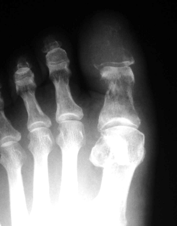
Foot radiograph of a 70-year-old man with intermittent sharp pain in his left first metatarsal.
This patient was a heavy smoker and had a dry cough. In the 2 months before presentation, he had experienced an unintentional 4 kg-weight loss. A radiograph showed an osteolytic lesion at the distal phalanx of his left first metatarsal (Figure 1). A bone scan showed increased tracer uptake at this site (Figure 2) and other parts of the left foot. A chest radiograph showed a mass in the right lower lobe of the lung, confirmed to be squamous cell lung cancer. An excisional biopsy of the phalangeal lesion revealed metastatic squamous cell carcinoma. The patient received palliative chemotherapy; he died 11 months after diagnosis.

Figure 1: Radiograph of left foot showing an osteolytic lesion (arrow) at the distal phalanx of the first metatarsal.

Figure 2: Bone scan showing increased tracer uptake at the left first metatarsal (arrow), ankle and heel.
Acrometastasis usually presents as a manifestation of widespread metastasis; however, in rare cases it can be the first sign of metastatic disease.1 In such cases it is often mistaken for an inflammatory or metabolic condition (e.g., gout, pseudogout, osteoarthritis, nondisplaced fractures) or a soft-tissue infection, which results in a delay in diagnosis, inappropriate therapy and inaccurate tumour staging.2 Fine-needle aspiration or biopsy for cytology allows differentiation of malignant and benign processes.
Footnotes
-
Competing interests: None declared.
In this issue

Article tools


Jump to section
Related Articles
Cited By...
- No citing articles found.
More in this TOC Section
Similar Articles
Collections

