


A 77-year-old woman was admitted to hospital with acute coronary syndrome and hypertensive crisis (i.e., a blood pressure of 210/110 mm Hg on admission). We administered acetylsalicylic acid, metoprolol and enoxaparin. One day later, after her blood pressure was stable and we had ruled out myocardial infarction, we performed an adenosine stress test. During the test, the patient became hypotensive and experienced nausea, confusion and abdominal pain.
She had a blood pressure of 70/40 mm Hg and a heart rate of 68 beats/min. She had no jugular venous distension. Her lungs were clear and she had a regular heart rhythm without murmurs, gallops or rubs. Her abdomen was tender over the right lower quadrant and she had a palpable 12 × 5 cm mass in this area with a positive Carnett sign (i.e., an increase in abdominal pain when the head and shoulders are lifted off the examination table).
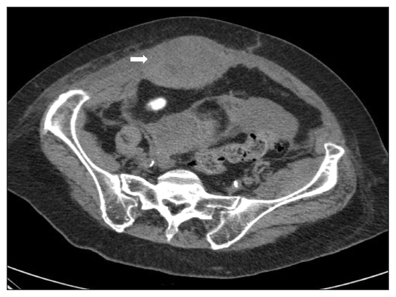
The patient’s cardiac troponin levels and an electrocardiogram were normal. Her hemoglobin level had dropped from 100 on admission to 71 (normal 129–158) g/L and her platelet count was 306 (normal 150–300) × 109/L. Prothrombin time and international normalized ratio were within normal range. A computed tomography scan of the abdomen showed a right rectus sheath hematoma associated with a pelvic hematoma (Figure 1). We immediately withheld antihypertensive agents and enoxaparin, performed fluid resuscitation and administered three units of packed red blood cells. The patient’s hemoglobin level normalized and she recovered uneventfully.

Figure 1: Computed tomography scan of the abdomen of a 77-year-old woman taking anticoagulants, showing a right-sided rectus sheath hematoma (arrow).
Rectus sheath hematoma is a rare but potentially life-threatening condition. In one series of rectus muscle masses, rectus sheath hematoma was the most common non-neoplastic cause (22%). 1 Although the condition appears to be an uncommon complication of anticoagulation, the incidence among patients receiving anticoagulant therapy is unknown. Risk factors for rectus sheath hematoma include advanced age, systemic anticoagulation (present in 69% of hematomas), intra-abdominal injections, strain of the abdominal wall, minor trauma and pregnancy. 2 Clinical manifestations include abdominal pain (84%), Fothergill sign (i.e., an abdominal mass that does not cross the midline and remains palpable when the rectus muscles are flexed), a substantial drop in hemoglobin, abdominal wall ecchymosis (present in 21% of hematomas) and a positive Carnett sign. 2,3 A positive Carnett sign suggests the origin of the pain is extra-abdominal rather than intra-abdominal. 3 The diagnosis can be confirmed by ultrasound or computed tomography scan of the abdomen.
Treatment of rectus sheath hematoma is usually expectant and may include fluid resuscitation, blood transfusions and management of pain. Uncommonly, intravascular embolization or surgery may be needed. Outcomes are usually favourable, although fatal outcomes have been reported. 4
Footnotes
-
This article has been peer reviewed.
Competing interests: None declared.
In this issue
Article tools


Jump to section
Related Articles
Cited By...
- No citing articles found.
More in this TOC Section
Similar Articles

