


- View this table:
Table 1.
- View this table:
Table 2.
- View this table:
Table 3.
Fig. 1: Transvaginal ultrasound image, showing intrauterine fluid collection without yolk sac or fetal pole: “pseudogestational sac” (arrow).
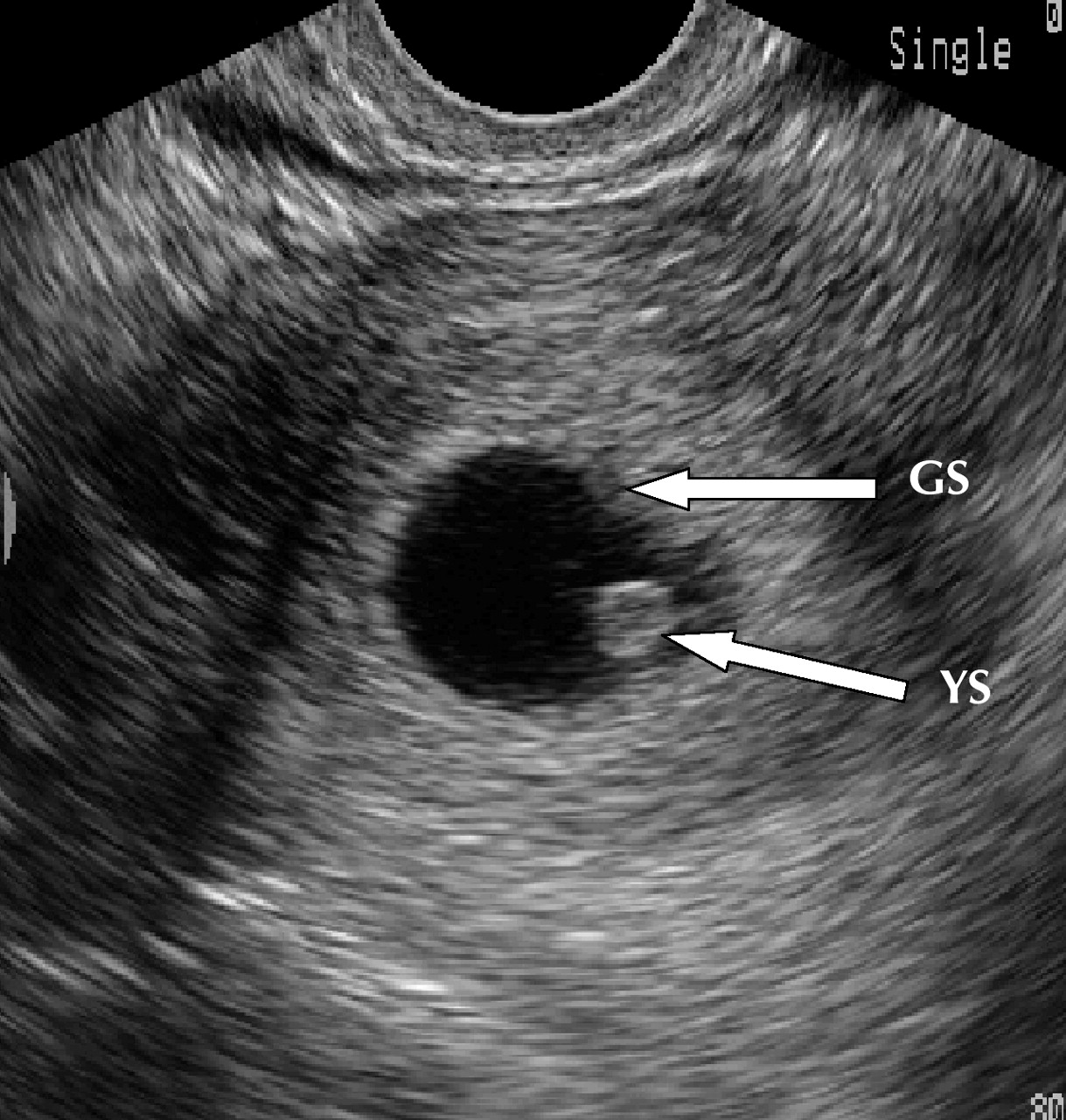
Fig. 2: Transvaginal ultrasound image, showing early intrauterine gestational sac (GS) with yolk sac (YS).
Fig. 3: Transvaginal ultrasound image, showing empty uterus and complex adnexal mass (ectopic pregnancy [EP]) separate from ovary.
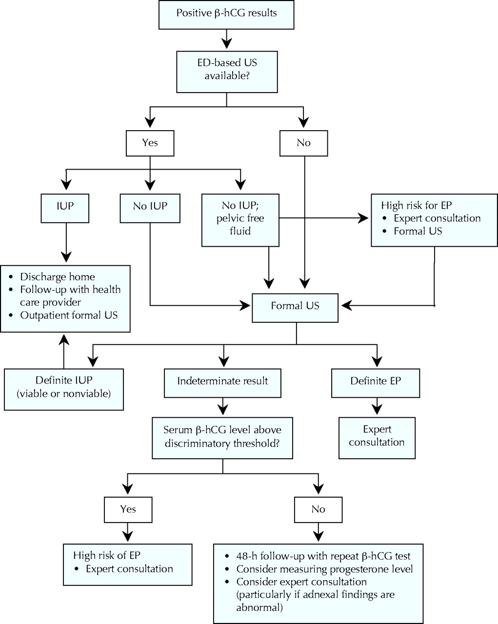
Fig. 4: Recommended approach to investigating first-trimester pain or bleeding in the hemodynamically stable patient in the emergency department (ED). β-hCG = β human chorionic gonadotropin, US = ultrasonography, IUP = intrauterine pregnancy, EP = ectopic pregnancy.
- View this table:
Table 4.


Article tools


Jump to section
Related Articles
Cited By...
- Point-of-care ultrasound for evaluation of vaginal bleeding or abdominal pain in early pregnancy: Use by family physicians following focused training and certification
- Ovarian vein thrombosis: a potential clinical consequence of ruptured ectopic pregnancy
- Hemoperitoneum in a Peritoneal Dialysis Patient: Ruptured Ectopic Pregnancy
- Impact of Accreditation Training for Residents on Sonographic Quality in Gynecologic Emergencies
- Cesarean Scar Pregnancy: Sonographic and Magnetic Resonance Imaging Findings, Complications, and Treatment
- Circulating MicroRNA miR-323-3p as a Biomarker of Ectopic Pregnancy
- Diagnosis and management of ectopic pregnancy
- Diagnosis and management of ectopic pregnancy
More in this TOC Section
Similar Articles
Collections

