


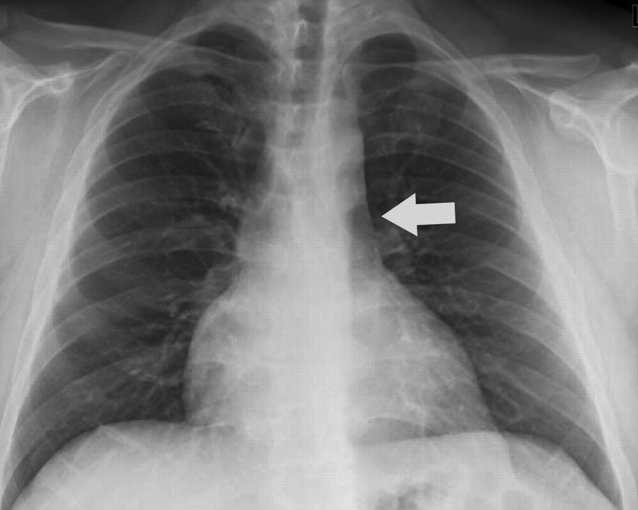
Fig. 1: Chest radiograph, posteroanterior view. The vascular shadow (arrow) in the upper mediastinum, extending from the clavicle along the left margin of the aortic arch, reveals the left-sided superior vena cava.
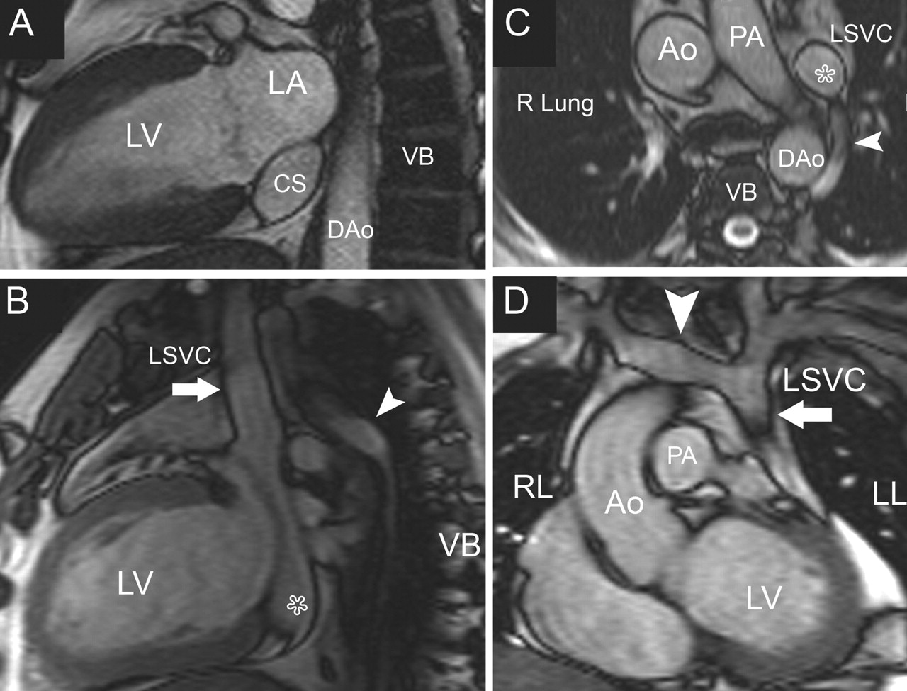
Fig. 2: Cardiovascular MRI, “bright blood” sequence. A. Scan of the cardiac 2-chamber plane: the coronary sinus is severely dilate. B. Thoracic sagittal plane: the left SVC (arrow) is connected to the cardiac sinus (star) and the left azygos vein (arrowhead) drains into the left SVC. C. Transverse plane through the upper thorax: the left SVC (star) and left azygos vein (arrowhead) are evident, whereas the right SVC is absent. D. Coronal thoracic plane: The innominate vein (arrowhead) connects the right jugular and subclavian veins to the left SVC (arrow). Ao = ascending thoracic aorta, DAo = descending thoracic aorta, LA = left atrium, LL = left lung, LSVC = left-sided superior vena cava, LV = left ventricle, PA = pulmonary artery, RL = right lung, SVC = superior vena cava, VB = vertebral body.
Fig. 3: The development of persistent left-sided superior vena cava (SVC). Left panel: The embryonic venous system, in which the superior and inferior caval veins (CVs) join into the common caval vein. At 8 weeks, the innominate vein links the superior CVs. Centre: The left superior CV normally obliterates distally to the innominate vein; the right-sided SVC develops from the right superior CV and part of the right common CV. The coronary sinus, which collects myocardial venous blood, develops from the left common CV. Right panel: A persistent left SVC connects to the coronary sinus. The right-sided SVC is absent because of persistence of the distal superior CV segment on the left and involution on the right. AntCV = superior caval veins, CCV = common caval vein, CS = coronary sinus, InV = innominate vein, LSVC = left-sided superior vena cava, PostCV = inferior caval veins.


Article tools


Related Articles
Cited By...
- No citing articles found.
More in this TOC Section
Similar Articles
Collections

