


Chewing betel quid or the combination of chewing betel quid and smoking cigarettes is associated with an increased risk of oral squamous cell carcinoma.1 The composition of betel quid varies with geographic location. In Taiwan betel quid is composed of areca nut (Areca catechu, an Asian tropical palm), slaked lime, and the inflorescence or leaf of Piper betle (an Asian climbing plant). The inflorescence of Piper betle contains high concentrations (15 mg/g fresh weight) of safrole, an essential oil used in cosmetics and as a food flavouring. Safrole is classified as a rodent hepatocarcinogen,2 and chewing betel quid may contribute to human exposure to this compound. The saliva of a person chewing betel quid contains on average 420 μmol/L of safrole.3
We describe a case of hepatocellular carcinoma in a Taiwanese man who had chewed betel quid for over 32 years; safrole-DNA adducts, a likely cause of liver carcinogenesis, were found in liver biopsy specimens.
Case
A 54-year-old man presented to hospital with an oral mass subsequently diagnosed as oral squamous cell carcinoma. His past medical history was unremarkable, and he had worked most of his life as a taxi driver. He admitted to heavy use of betel quid (about 30 betel quids daily over 32 years). In addition he had smoked 1.5 packs of cigarettes daily for the same period. He consumed alcohol only on social occasions and then only in moderate amounts. Physical examination revealed a nontender firm mass in the right upper quadrant of the abdomen. Liver echography and CT revealed a hypervascular tumour mass about 4 cm in diameter located in the lateral aspect of the right hepatic lobe. The results of liver function tests included alanine aminotransferase 32 (normally 5 to 35) U/L, aspartate aminotransferase 28 (normally 5 to 30) U/L, alkaline phosphatase 52 (normally 25 to 100) U/L and α-fetoprotein 67 (normally less than 6) μg/L. The patient was not infected with hepatitis B or C virus (positive for antibodies to hepatitis B surface antigen and negative for both hepatitis B surface antigen and hepatitis C surface antigen).
Liver biopsy (Fig. 1) showed classic hepatocellular carcinoma. Using the nuclease P1-enrichment version of the 32P-postlabelling technique,4 we detected safrole-DNA adducts as a single spot on the autoradiogram (Fig. 2). Similar safrole-DNA adducts were seen in tissue samples from the oral squamous cell carcinoma and in peripheral blood leukocyte samples.5 The level of safrole-DNA adduct detected was 22.5 adducts per 108 nucleotides in liver, 7.1 adducts per 108 nucleotides in the oral squamous cell carcinoma and 0.8 adducts per 108 nucleotides in peripheral blood leukocytes. The profile and location of the safrole-DNA adduct were similar to those of adduct found in HepG2 cells treated with 1′-hydroxysafrole; the adduct has been identified as N2-(trans-isosafrole-3′-yl)2′-deoxyguanosine.[5, 6] In parallel studies using similar tissues obtained from 6 people who had hepatocellular carcinoma or oral squamous cell carcinoma and who did not chew betel quid, we were unable to detect the safrole-DNA adduct (unpublished data).

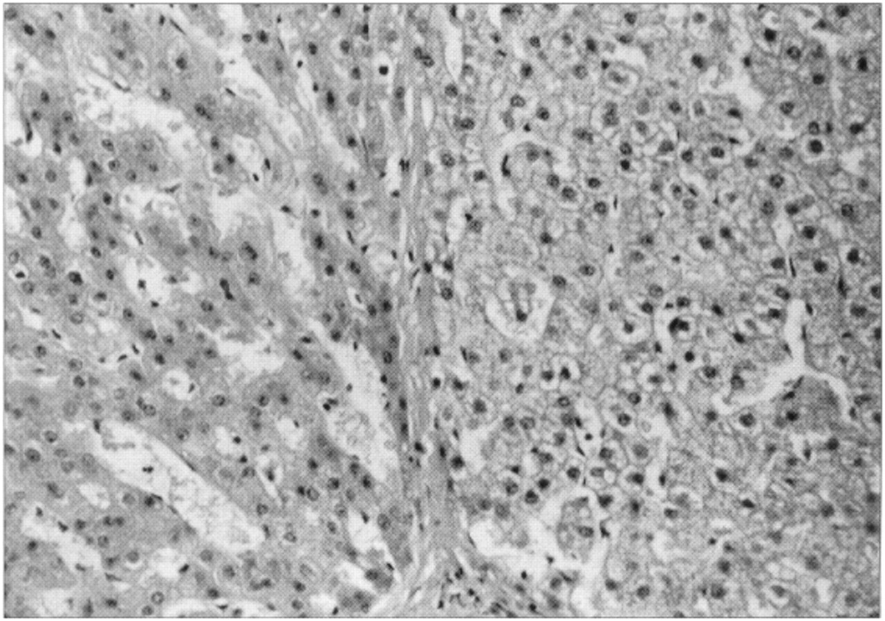
Fig. 1: Microscopic image of a liver section stained with hematoxylin and eosin exhibits well-differentiated hepatocellular carcinoma. The tumour is localized on the left side of the image and is separated from normal liver parenchyma (at right) by a thin layer of fibrous connective tissue. The tumour cells are arranged in a sinusoid pattern. The cytoplasm has a clear or ground-glass appearance. Slight cellular or nuclear pleomorphism is evident.

Fig. 2: Autoradiograms of polyethyleneimine-cellulose thin layer chromatography maps of 32P-labelled digests of DNA (autoradiography performed with Kodak Biomax MR film for 3 hours at −70°C). Left: DNA from HepG2 cells treated with 400 μmol/L 1′-hydroxysafrole for 24 hours. One safrole-DNA adduct visualized as a black spot can be clearly seen in the bottoom left corner of this autoradiogram. Right: DNA from liver biopsy specimen. A safrole-DNA adduct can be seen at the bottom left corner of this autoradiogram.
Comments
Carcinogen-DNA adducts represent chemical modifications to the genetic material. They usually arise from the bioactivation of a carcinogen, which then reacts with the DNA. The damage caused by adducts is central to theories of chemical carcinogenesis and is considered a necessary prerequisite for gene mutation and tumour formation.7 Studies in mice have shown that safrole-induced liver carcinogenesis is correlated with the formation of safrole-DNA adducts.8 This type of adduct is created through cytochrome-P450-mediated formation of 1′-hydroxysafrole; this compound is sulfonated to become an unstable sulfuric acid ester, which then forms the stable safrole-DNA adducts.9
Although these studies do not prove that the safrole in betel quid caused our patient's hepatocellular carcinoma, the findings are suggestive. We found safrole-DNA adducts in the nucleotides of the biopsy specimen from the hepatocellular carcinoma. The distribution of these adducts was similar to that found in safrole-treated mice: highest in the liver and lower in other tissues (the level in peripheral blood leukocytes was only 1/51 the level in the liver). Our preliminary observations indicate that human tissue harbours the potential to bioactivate the safrole in betel quid to its corresponding DNA adducts, particularly in the liver. This study is the first to show the presence of stable safrole-DNA adducts in hepatocellular carcinoma and oral squamous cell carcinoma in a heavy betel quid user.
Competing interests: None declared.
Acknowledgments
Seek an opinion, share an insight or wax poetic with fellow members of the CMA at www.cma.ca/discussion_groups
Footnotes
-
This article has been peer reviewed.
Reprint requests to: Dr. Tsung-Yun Liu, Department of Medical Research, Veterans General Hospital - Taipei, Shih-Pai, Taipei, 11217, Taiwan, Republic of China; fax 886 2 2875 1562; [email protected]
In this issue
Article tools


Jump to section
Related Articles
Cited By...
More in this TOC Section
Similar Articles

