


A 46-year-old man presented with a 2-year history of asymptomatic erythematous annular plaques on his upper chest (Figure 1), neck, upper back and dorsum of hands (Figure 2). The lesions started as small papules, which slowly expanded centrifugally to form annular plaques with atrophic centres. Routine laboratory, antinuclear antibody and extractable nuclear antigen tests were negative or within normal limits. Histopathologic examination was consistent with actinic granuloma (Appendix 1, available at www.cmaj.ca/lookup/suppl/doi:10.1503/cmaj.190120/-/DC1). We advised our patient about sun protection and prescribed oral prednisolone 30 mg/d, tapering by 5 mg/wk until a dose of 5 mg/d was reached, which we continued for 3 months. At that point, our patient’s lesions had flattened, and no new lesions had developed.
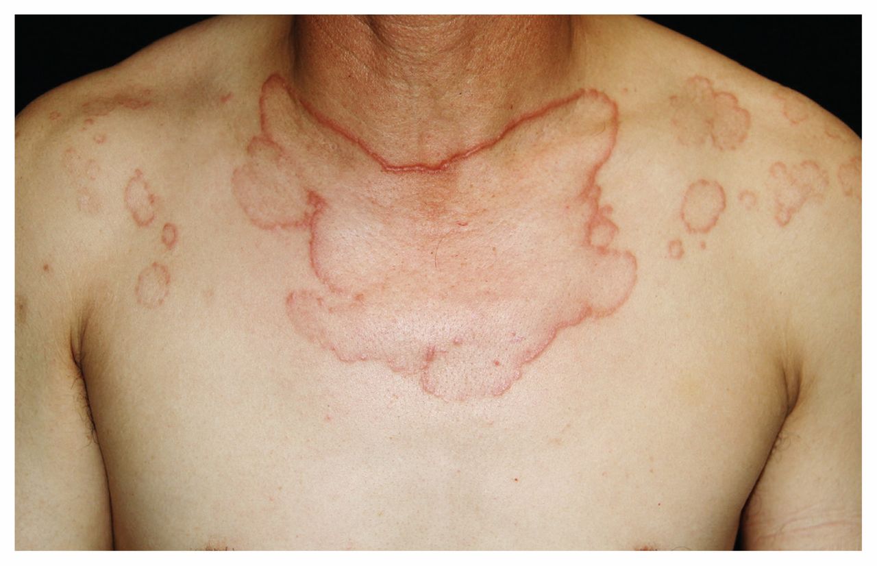
Multiple asymptomatic erythematous annular plaques with atrophic centres on the upper chest of a 46-year-old man.

Asymptomatic erythematous annular plaques with atrophic centres on the dorsum of the patient’s hands.
Actinic granuloma is a chronic, benign granulomatous condition that affects middle-aged people who have a history of intense sun exposure. It typically presents as annular plaques with raised erythematous borders and atrophic centres on sunexposed areas. Lesions can initially occur as erythematous papules and then gradually expand centrifugally and coalesce into asymptomatic annular or linear plaques with elevated borders. Histopathologic examination shows granulomatous infiltration of the skin with histiocytes surrounded by multiple multinucleated giant cells containing degenerating elastic fibres.1
It is believed that a cell-mediated autoimmune response against damaged elastic fibres elicits the granulomatous inflammation.1 When actinic granuloma–like lesions are located in unexposed areas, the rash is described as annular elastolytic giant cell granuloma. The differential diagnosis includes tinea corporis, sarcoidosis, subacute lupus erythematosus, granuloma annulare and other infectious granulomatous diseases, but these can be distinguished readily on histopathologic features and laboratory findings. Treatments including chloroquine, intralesional or systemic steroids, cyclosporine, isotretinoin, acitretin and laser treatment have been tried with variable success.2,3
Footnotes
Competing interests: None declared.
This article has been peer reviewed.
The authors have obtained patient consent.
In this issue

Article tools


Related Articles
Cited By...
- No citing articles found.
More in this TOC Section
Similar Articles
Collections

