


Article Figures & Tables
Figures
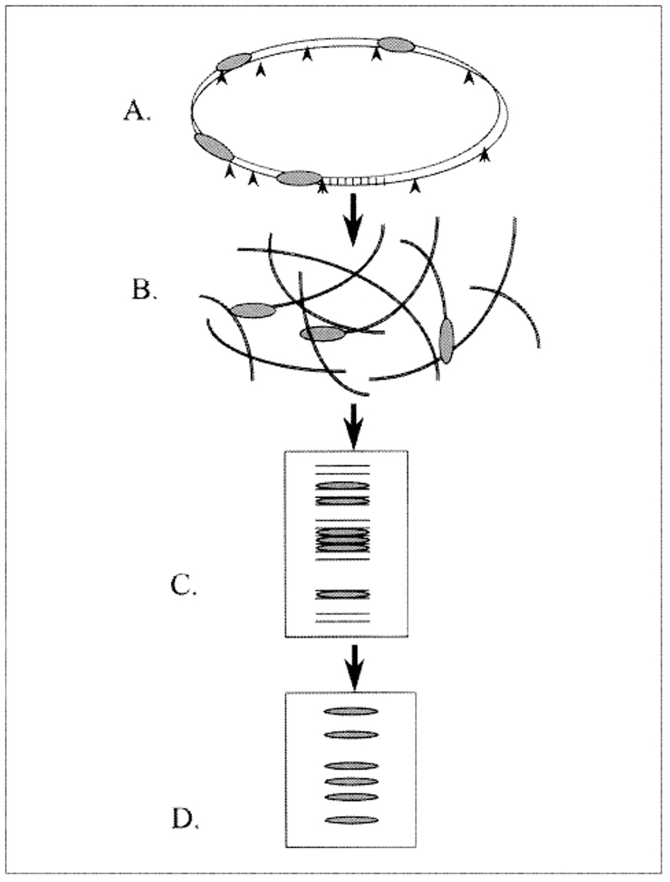
Fig. 1: Genetic fingerprinting of Mycobacterium tuberculosis isolates: (A) Restriction enzymes cleave chromosomal DNA at restriction sites (arrowheads). (B) Some DNA fragments contain IS6110 (repetitive sequences of base pairs, represented as shaded ellipses). (C) The fragments are separated according to size by gel electrophoresis. (D) Fragments containing IS6110 hybridize to the specific radioactive probe, which produces a characteristic banding pattern (fingerprint) for each isolate.
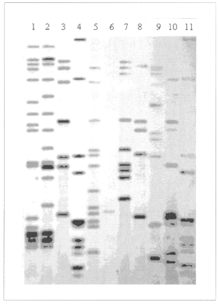
Fig. 2: Autoradiograph, showing IS6110-based DNA fingerprints of M. tuberculosis isolates. Lanes 4 and 11 represent molecular weight ladders that provide information about the length of DNA fragments. The other 9 lanes contain whole genomic DNA of different isolates. Lane 6 contains a one-copy strain whose fingerprint is of low resolution; to investigate links between such a strain and others with the same one-copy fingerprint, secondary typing with another marker system is usually performed.

In this issue
Article tools


Jump to section
Related Articles
Cited By...
More in this TOC Section
Similar Articles
Collections

