


Article Figures & Tables
Figures
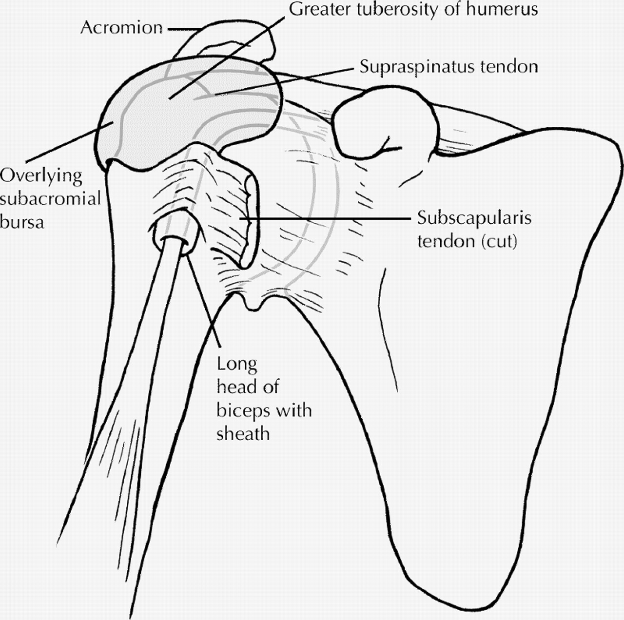
Fig. 1: Anatomy of the shoulder joint showing rotator cuff tendons, subacromial bursa and biceps tendons. Photo by: Lianne Friesen
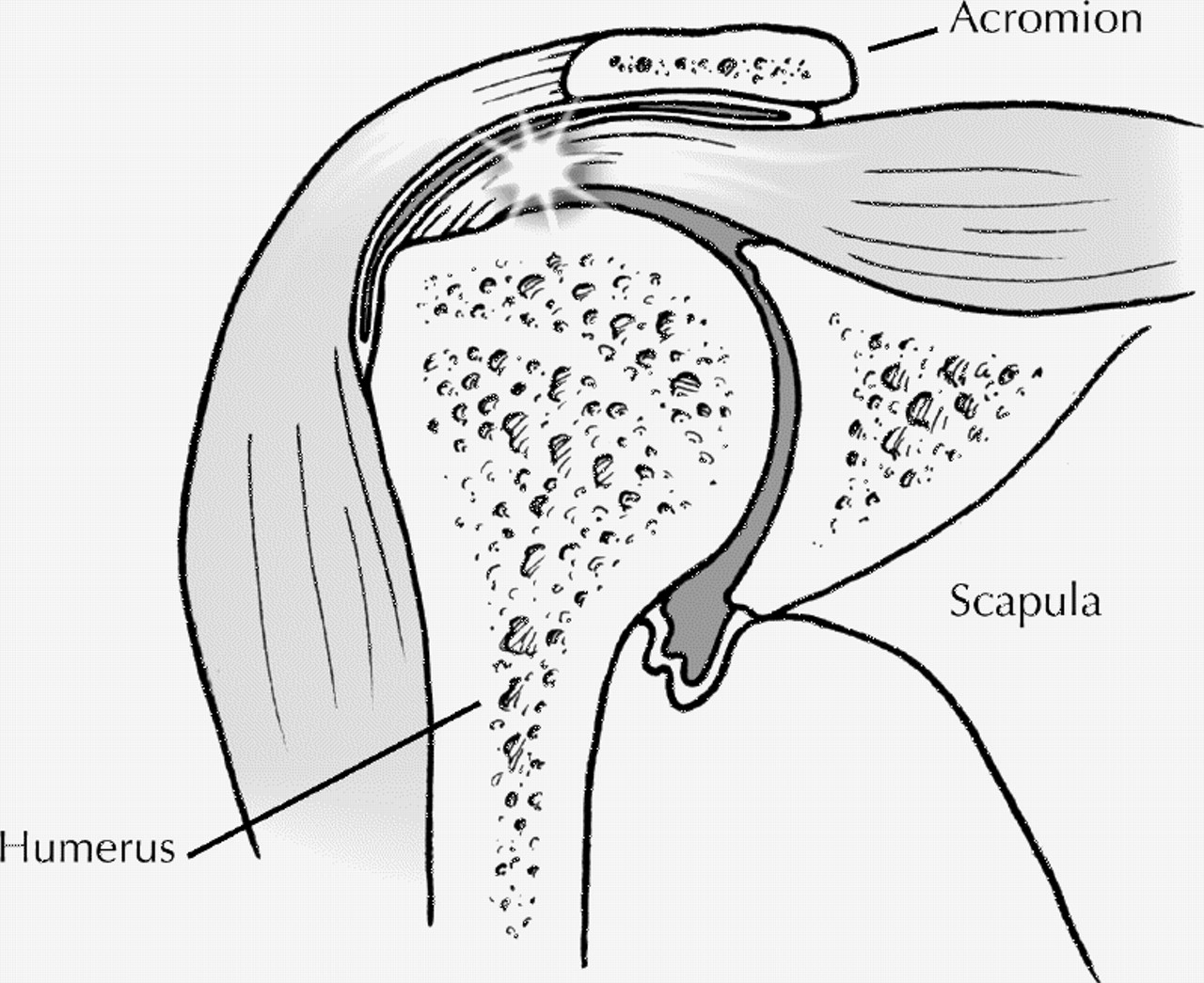
Fig. 2: Mechanism of impingement sign: inflamed region of supraspinatus tendon and/or adjacent bursa. Photo by: Lianne Friesen
Figure 2. Continued.
Figure 2. Continued.
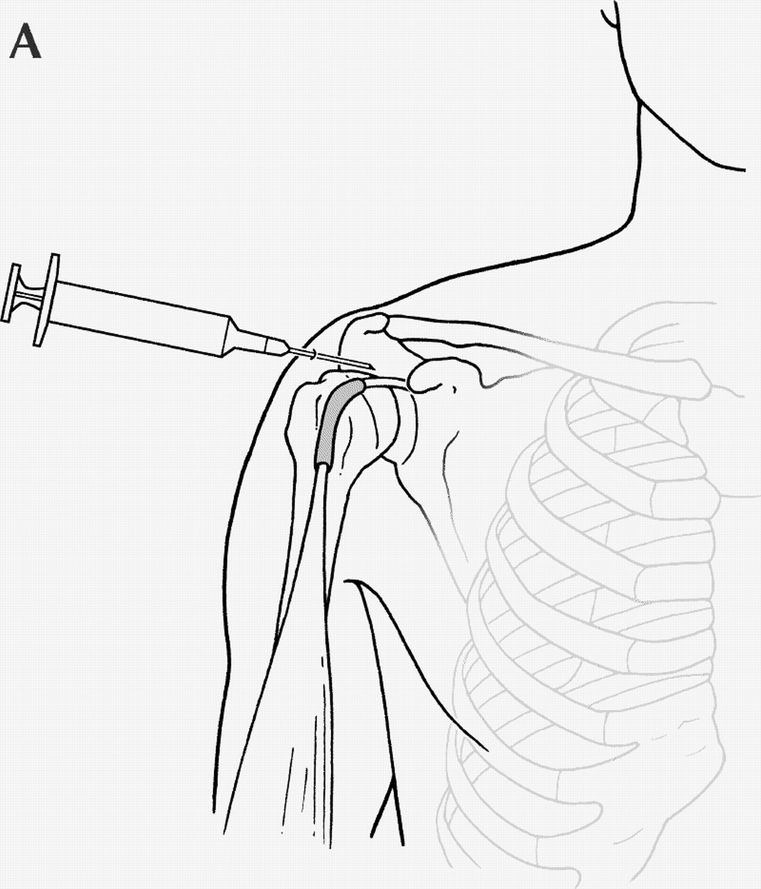
Fig. 3: Periarticular shoulder injection (A); infiltration of the tendon sheath of the long head of the biceps (B). Photo by: Lianne Friesen
Figure 3. Continued.
Fig. 4: Tendons involved in de Quervain's tenosynovitis. Photo by: Lianne Friesen


Tables
Table 1.


In this issue
Article tools
Respond to this article


Jump to section
Related Articles
Cited By...
More in this TOC Section
Similar Articles

