


Article Figures & Tables
Figures
Fig. 1: Global distribution of malaria. Drug resistance is represented by the shaded areas (see legend). This map is intended as a visual aid only; online sources of country-specific malaria risk are provided in “Additional Resources.” Reproduced, with permission, from the Committee to Advise on Tropical Medicine and Travel, Health Canada. Canadian recommendations for the prevention and treatment of malaria among international travellers — 2003. Can Commun Dis Rep 2004;30(Suppl 1). In press.
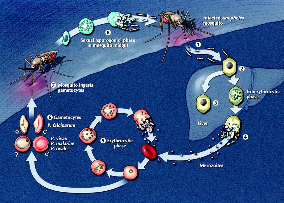
Fig. 2: The plasmodia life cycle. The human (asexual) stage of the life cycle begins with the exoerythrocytic phase. When an infected mosquito bites a human, sporozoites in the mosquito's saliva enter the bloodstream (1). The sporozoites travel to the liver, where they invade hepatocytes (2); over a period of up to 4 weeks, the infected hepatocytes mature into schizonts. In Plasmodium vivax and P. ovale infections only, some schizonts may remain dormant as hypnozoites (3) for weeks to years before causing clinical relapses. With schizont rupture, merozoites are released into the bloodstream (4). In the erythrocytic phase, merozoites invade erythrocytes and either undergo an asexual cycle of reproduction (5) or develop into nonmultiplying sexual forms (gametocytes) (6). These gametocytes are crucial for perpetuating the life cycle, as they are ingested by a feeding mosquito (7) and undergo sexual reproduction within the mosquito midgut; thousands of infective sporozoites (8) are produced, which then migrate to the salivary glands, ready to initiate another life cycle. Photo: Lianne Friesen and Nicholas Woolridge
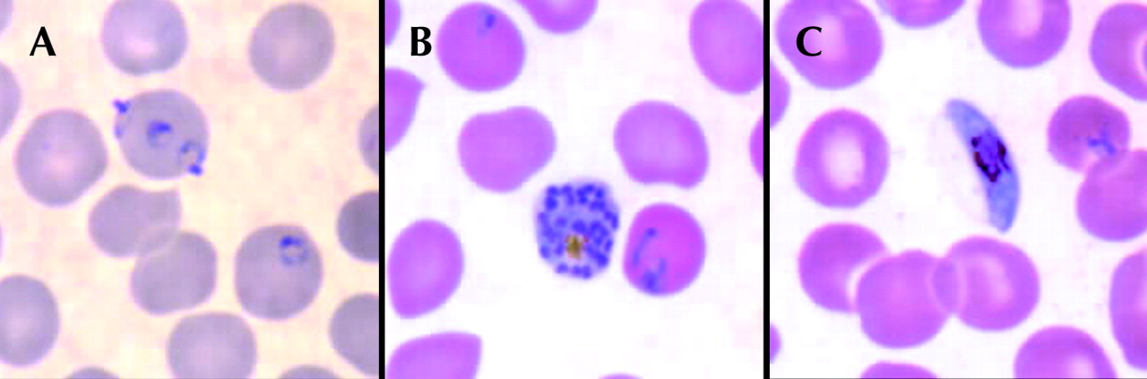
Fig. 3: Stages in the life cycle of Plasmodium falciparum. A: Ring forms (early trophozoites). B: Mature schizont, rarely seen in peripheral blood smears because of microvascular sequestration. C: Gametocyte, demonstrating the classic banana shape. Source: Division of Parasitic Diseases, US Centers for Disease Control and Prevention, Atlanta. Photo: CDC


Tables
Table 1.

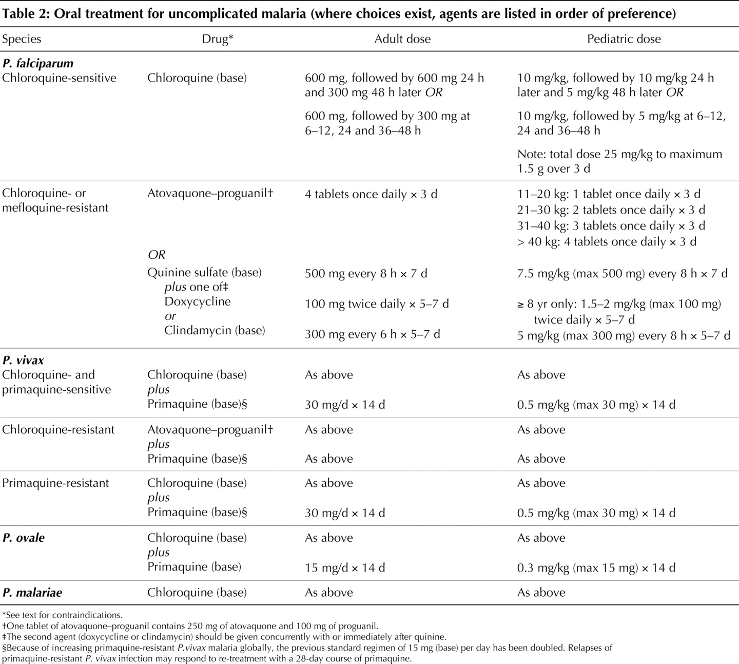
Table 2.
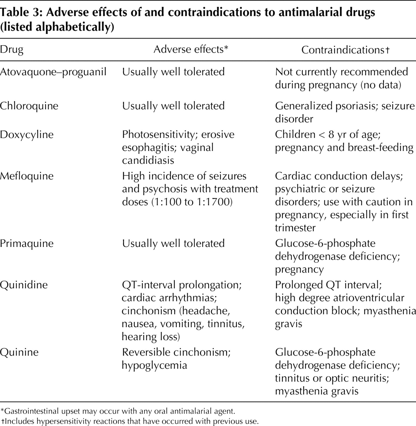
Table 3.
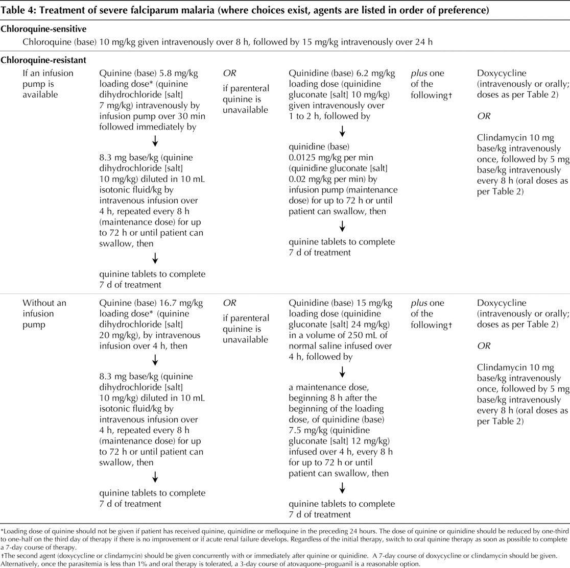
Table 4.
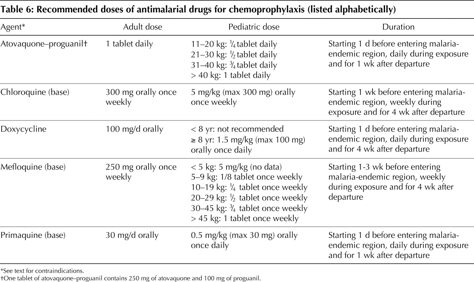
Table 6.
Table 5.


In this issue
Article tools


Jump to section
Related Articles
Cited By...
- No citing articles found.
More in this TOC Section
Similar Articles
Collections

