


Article Figures & Tables
Figures
Fig. 1: CT scan of the chest showing (A) nodular density in lateral segment of right lower lobe and (B) cavitated areas with surrounding enhancement extended to the chest wall.
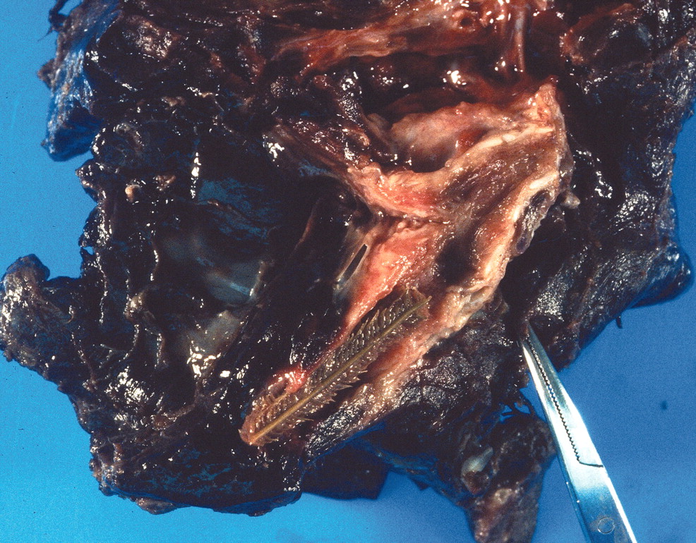
Fig. 2: Right lower lobectomy specimen. The embedded foreign body can be seen in the dissected lobar bronchus.

In this issue
Article tools
Respond to this article


Jump to section
Related Articles
Cited By...
More in this TOC Section
Similar Articles
Collections

