


Article Figures & Tables
Figures
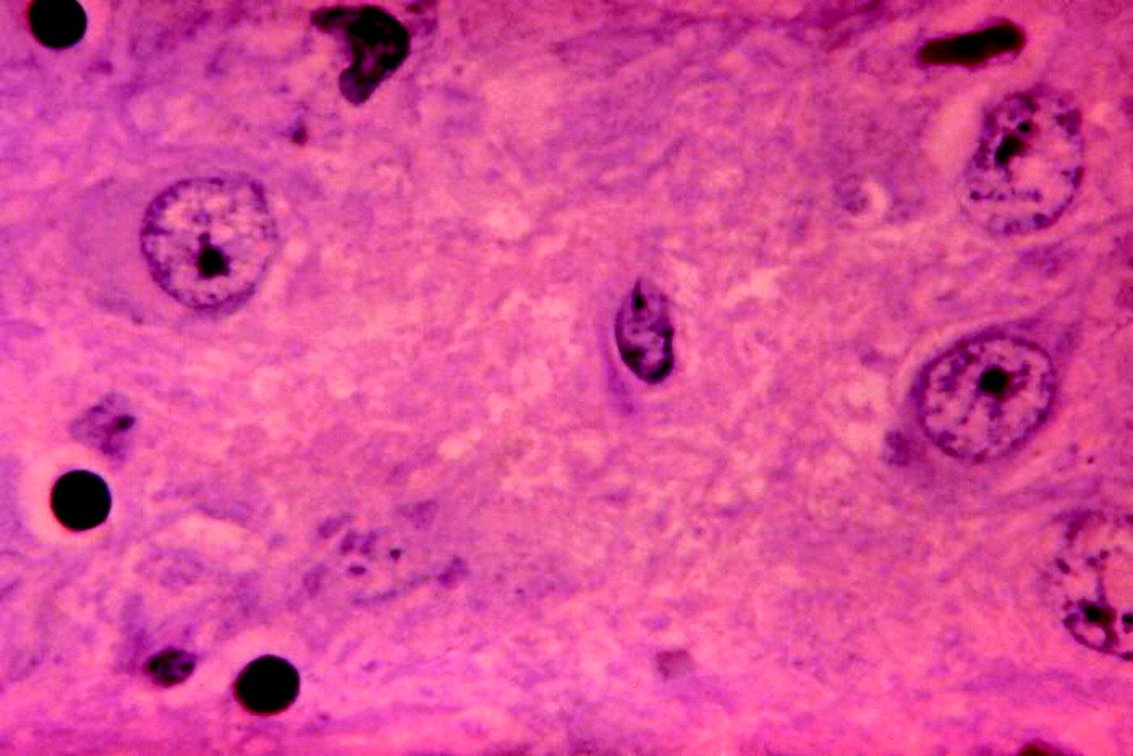
Fig. 1: Micrograph showing histopathologic changes associated with rabies encephalitis. The pathognomonic Negri bodies are cellular inclusions found most frequently in the pyramidal cells of Ammon's horn and the Purkinje cells of the cerebellum. Photo: CDC/Dr. Daniel P. Perl
In this issue
Article tools
Respond to this article


Jump to section
Related Articles
Cited By...
- No citing articles found.
More in this TOC Section
Similar Articles
Collections

