


Article Figures & Tables
Figures
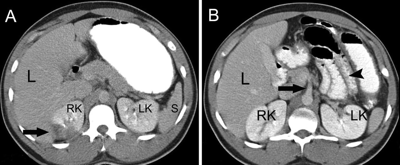
Fig. 1: Abdominal CT scans with contrast medium. Left: Hypodense lesion in right kidney (arrow), indicating infarction. Right: Filling defect in lumen of superior mesenteric artery (arrowhead), consistent with thrombus or thromboembolus, and edema of wall of small intestine (arrow). L = liver, RK/LK = right/left kidney, S = spleen.
Fig. 2: Intraoperative transesophageal echocardiogram, showing mobile thrombus (arrow) 1.5 ∞ 2 cm in size in descending thoracic aorta near origin of left subclavian artery.


In this issue
Article tools
Respond to this article


Jump to section
Related Articles
Cited By...
- No citing articles found.
More in this TOC Section
Similar Articles
Collections

