


Article Figures & Tables
Figures
Fig. 1. Images of coronary arteries, revealed with intravascular ultrasonography (IVUS). A: A normal artery. The layers of the arterial wall, from innermost to outermost, are the intima, media and adventitia. The intima is an endothelial cell layer where atheroma accumulates; the external boundary of this layer is the internal elastic membrane. The media, composed of smooth muscle cells, elastin and collagen, is encircled by the external elastic membrane. The adventitia is mainly composed of fibrous tissue. In young people free of atherosclerotic disease, the 3 layers are difficult to see as separate structures because the media and intima are smaller than the resolution of IVUS (< 100 mm). B: Mixed plaque (fibrotic and calcific) with a dissection of the media at the „6 o'clock” position. C: In-stent restenosis occluding the lumen. D: In-stent restenosis without angiographically significant occlusion.
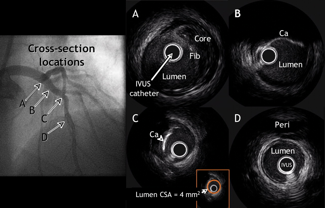
Fig. 2: The upper-left panel is a coronary angiographic image of clinically nonsignificant obstructive disease of the left anterior descending artery. Lettered arrows identify the locations of 4 IVUS cross-sections, as follows. A: In an area that appeared normal with angiography, a nonobstructive plaque is revealed containing a necrotic or lipid core covered by more echogenic tissue (Fib). B: In a section that likewise appeared normal with angiography, a calcified plaque can be seen. C: A severely calcified plaque with a minimum lumen cross-sectional area (CSA) of 4 mm2 (see inset). D: An IVUS cross-section showing a normal distal reference, the pericardium (Peri).
Fig. 3: Three months after insertion of a drug-eluting stent for in-stent restenosis in the middle segment of the left anterior descending artery, this patient was readmitted to hospital because of angina. Angiography showed a hazy lesion within the stent. IVUS imaging, however, did not reveal any notable intimal hyperplasia.
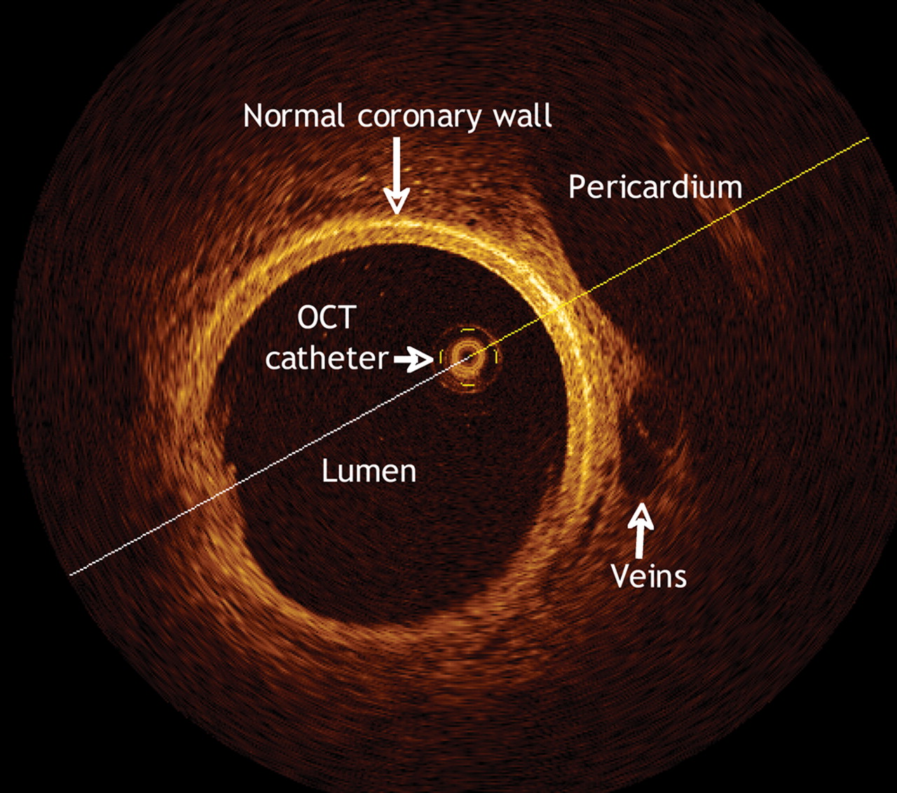
Fig. 4: Optical coherence tomographic (OCT) scan of a pig's normal coronary artery. The high resolution of the image is apparent, particularly in the details of the pericardium, veins and the coronary-artery walls.
Fig. 5: This patient experienced recurrent angina a year after undergoing percutaneous coronary intervention (PCI). Cardiac CT revealed a new critical stenosis (arrows) in the middle of the right coronary artery, which was confirmed by invasive angiography and subsequently treated with a second PCI. The 3-dimensional reconstruction (bottom) shows the stent clearly.
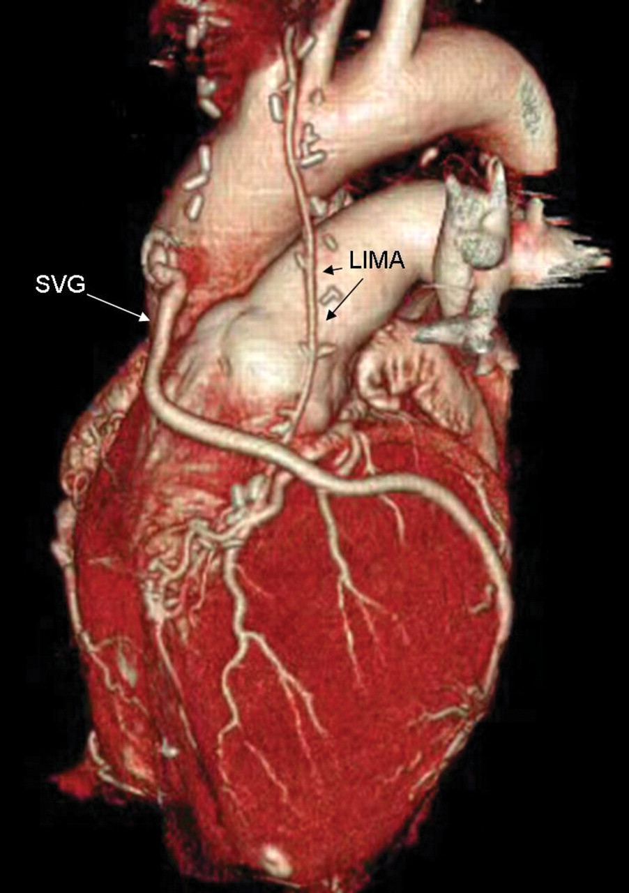
Fig. 6: A 3-dimensional reconstruction image showing patent bypass grafts in the left internal mammary artery (LIMA) and saphenous vein (SVG), by volume rendering. The entire data set for the image was acquired during a single 30-second breath-hold by the patient.
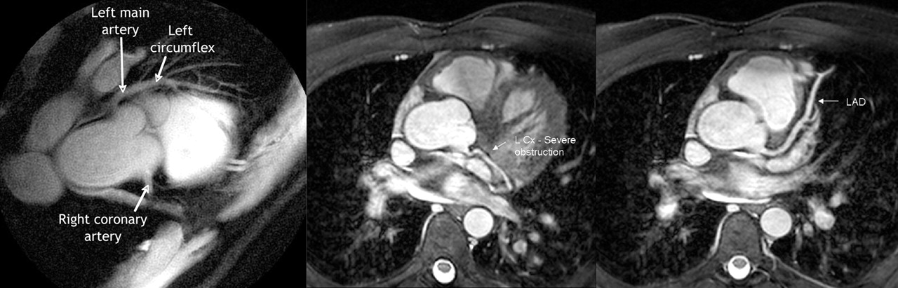
Fig. 7: MRIs of the heart of 1 patient. Left panel: the right coronary artery (showing proximal obstruction) and the left main and left anterior descending coronary arteries (no notable obstruction). Middle panel: the left circumflex (L Cx) of the coronary artery, with a proximal lesion. Right panel: the left anterior descending artery (LAD), with no lesions visible.


Tables
Table 1.

Table 2.
Table 3.


In this issue
Article tools


Jump to section
Related Articles
Cited By...
More in this TOC Section
Similar Articles

