


Article Figures & Tables
Figures
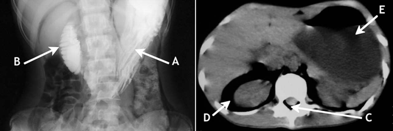
Fig 1: Left: Barium study, showing dilatation of the stomach (A) and the second and third portions of the duodenum, and obstruction of the duodenum (B). Right: CT scan of cervical spine, chest and abdomen, showing subcutaneous emphysema, pneumomediastinum, spinal epidural emphysema (C), retroperitoneal emphysema (D) and dilated stomach (E).
In this issue
Article tools
Respond to this article


Jump to section
Related Articles
Cited By...
- No citing articles found.
More in this TOC Section
Similar Articles
Collections

