


Article Figures & Tables
Figures
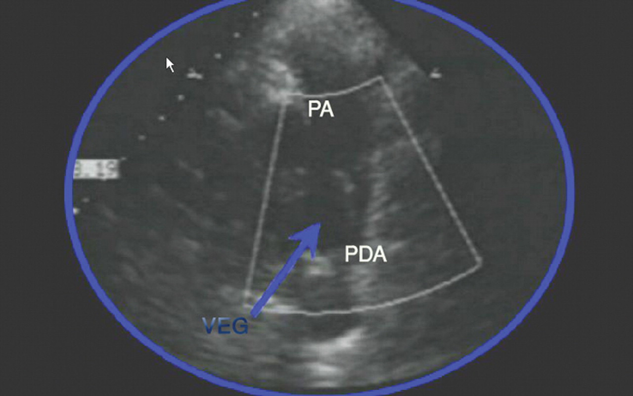
Fig. 1: Transthoracic echocardiogram showing a mobile mass of vegetation (VEG) at the left pulmonary artery (PA). PDA = patent ductus arteriosus.
Fig. 2: Schematic presentation of auscultatory findings in a patent ductus arteriosus. S1 and S2 denote the first and second heart sounds. The continuous murmur starts after S1, peaks near S2 and declines during diastole. The second heart sound may be masked by the murmur.


Article tools
Respond to this article


Related Articles
Cited By...
- No citing articles found.
More in this TOC Section
Similar Articles
Collections

