


What's your call?
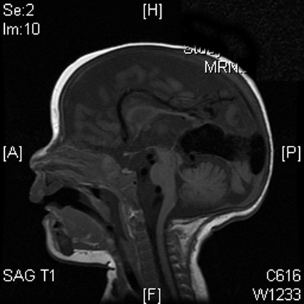
Figure. Sagittal T1-weighted cranial MRI scan of a 3-month-old boy who presented to the emergency department in congestive heart failure and with a harsh cranial bruit.
A large aneurysmal dilatation of the vein of Markowski and vein of Galen was noted on the cranial MRI. It was seen left of the midline, with marked mass effect, multiple fistulae and enlargement of the left middle-, left anterior-and posterior-cerebral arteries. MR venography showed a dilated venous pouch that drained into the falcine vein and posterior sagittal sinus (Fig. 1).

Fig. 1: Magnetic resonance venogram, showing a dilated venous pouch draining into the falcine vein and posterior sagittal sinus.
Vein of Galen malformations are rare congenital vascular abnormalities resulting in a direct communication between the cerebral arteries and the deep draining veins of the posterior cerebral fossa. The malformation develops between weeks 6 and 11 of fetal development as a persistent embryonic prosencephalic vein of Markowski, which drains into the vein of Galen. These veins become enlarged and aneurysmal. Vein of Galen malformations are usually diagnosed on antenatal ultrasonography in the third trimester. They can be associated with other conditions, including atrial septal defects, patent ductus arteriosis and pseudo-aortic coarctation (Pediatr Radiol 1997;27:501-13). Treatment usually involves endovascular embolization.
In this issue
Article tools


Jump to section
Related Articles
Cited By...
- No citing articles found.
More in this TOC Section
Similar Articles
Collections

