


Article Figures & Tables
Figures
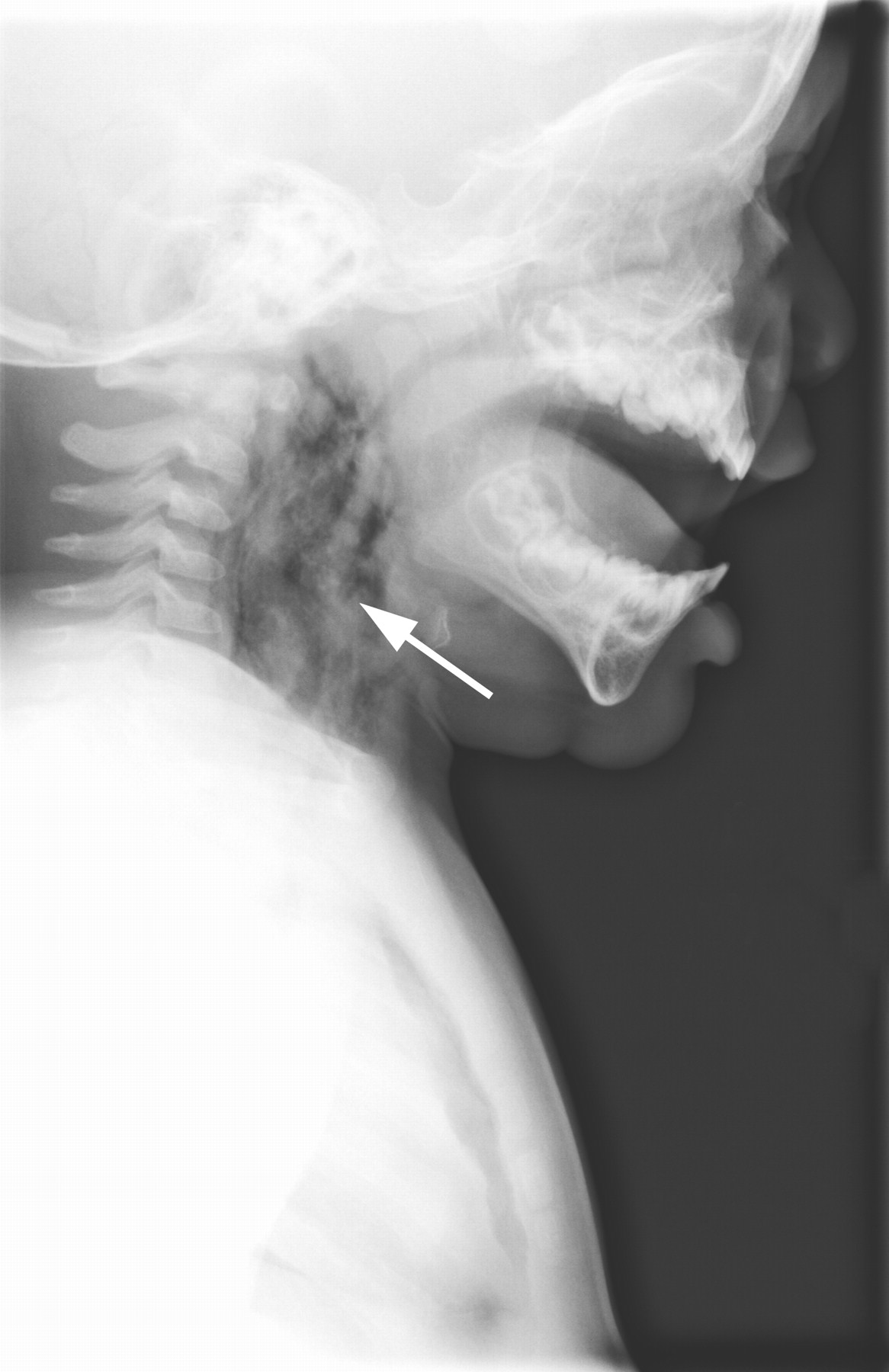
Figure 1: Neck radiograph showing diffuse subcutaneous emphysema (arrow) consistent with a puncture wound.
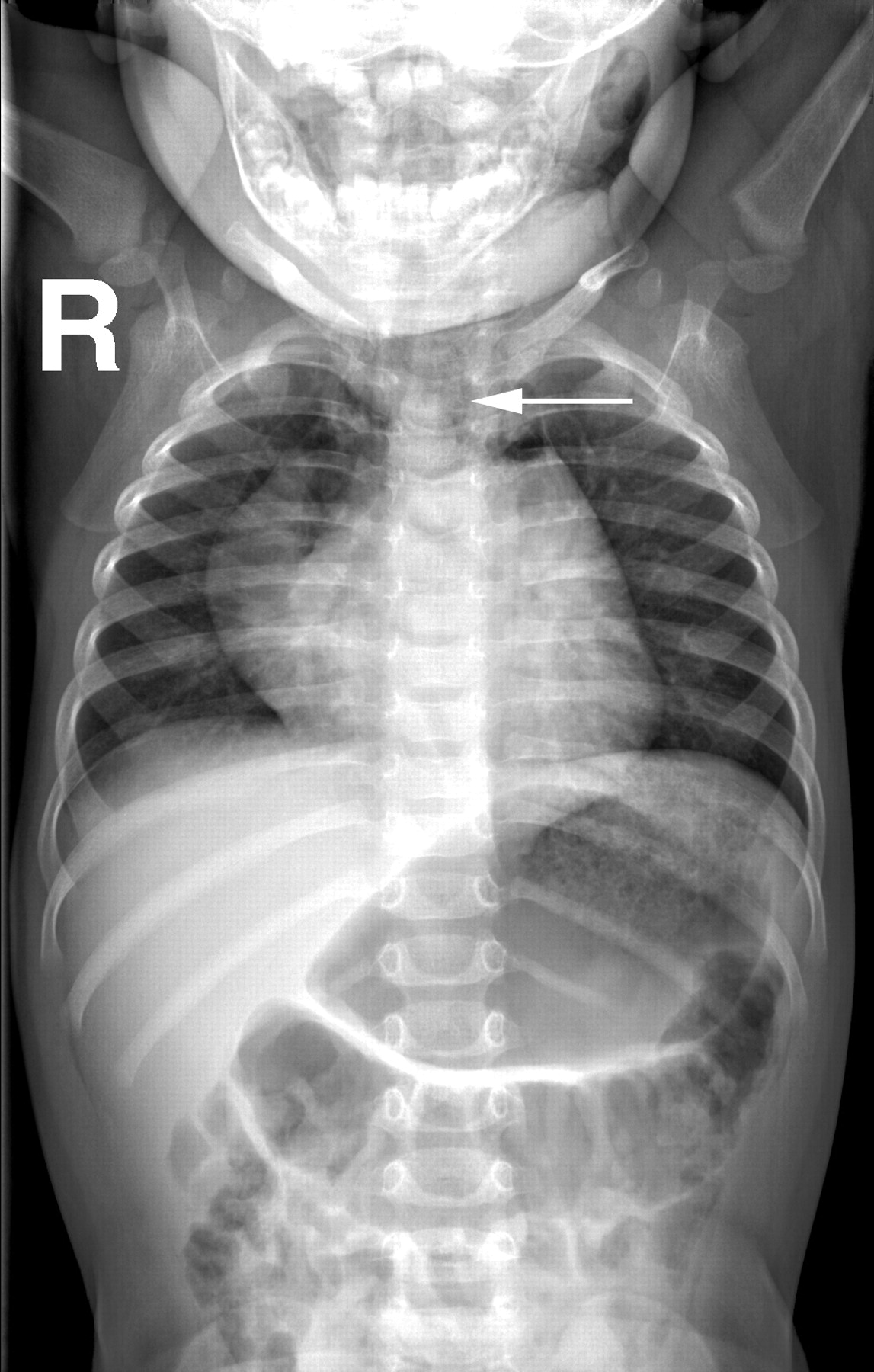
Figure 2: Chest radiograph showing emphysematous collections (arrow) along the fascial planes of the neck.
Figure 3: Computed tomography scan showing extensive emphysematous collections surrounding the carotids (arrow).


In this issue
Article tools
Respond to this article


Jump to section
Related Articles
Cited By...
More in this TOC Section
Similar Articles
Collections

