


Article Figures & Tables
Figures
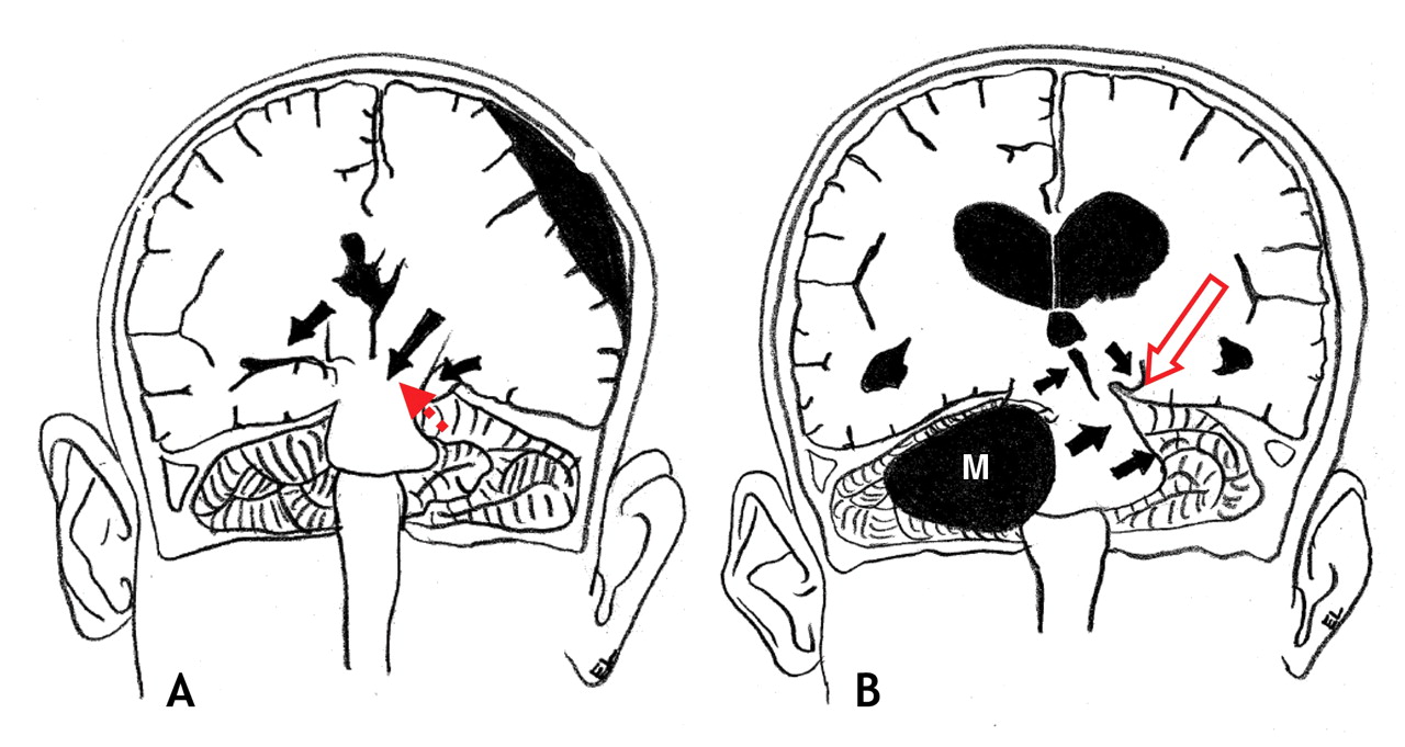
Figure 1: (A) Schematic coronal section of the brain showing large (supratentorial) right subdural hematoma causing ipsilateral transtentorial herniation, which has resulted in compression of the contralateral cerebral peduncle (broken, red arrow). This led to ipsilateral (right-sided) weakness. (B) Schematic coronal section of the brain showing (infratentorial) right meningioma (M) causing upward and leftward displacement of the midbrain and cerebral peduncles, which has resulted in compression of the left cerebral peduncle and corticospinal tracts against the tentorium cerebelli (open arrow), which contributed to the ipsilateral (right-side) weakness. In both figures, the solid arrows indicate the direction of shift due to the mass effect, either from the subdural hematoma or from the tumour.
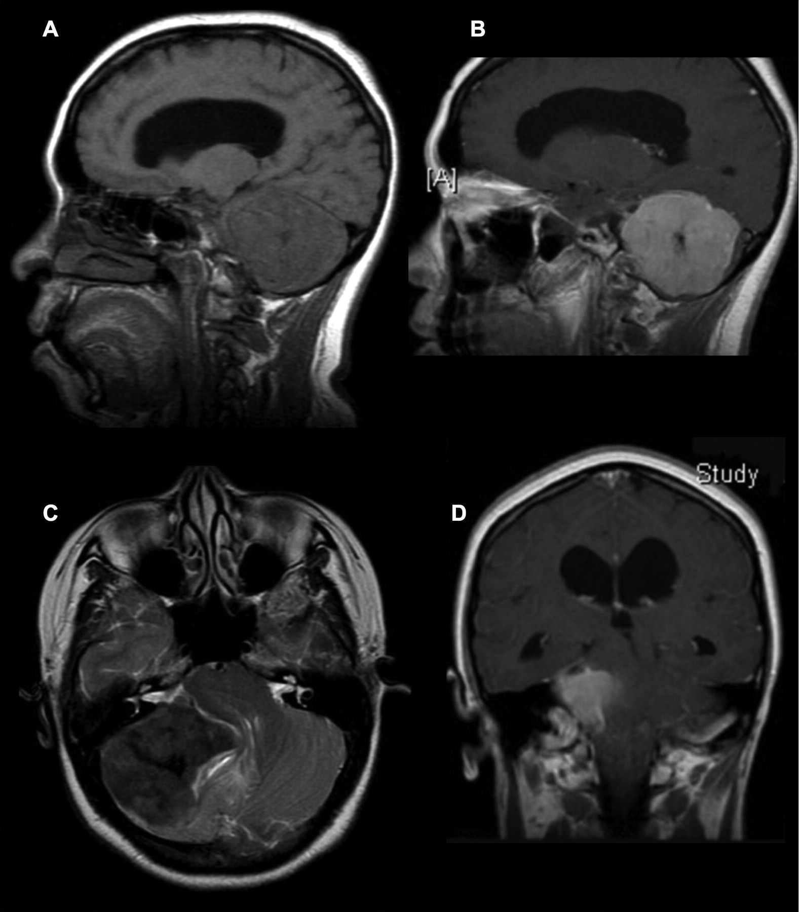
Figure 2: Sagittal T1-weighted MRI, without (A) and with (B) gadolinium contrast agent. Transverse T2-weighted (C) and coronal T1-weighted (D) images, both with gadolinium contrast agent, show 5 × 5.4 × 6 cm meningioma. The infratentorial mass is evident in Figure D, pushing the brain stem upward against the tentorium cerebelli at Kernohan's notch.


In this issue
Article tools


Jump to section
Related Articles
Cited By...
- No citing articles found.
More in this TOC Section
Similar Articles

