


Article Figures & Tables
Figures
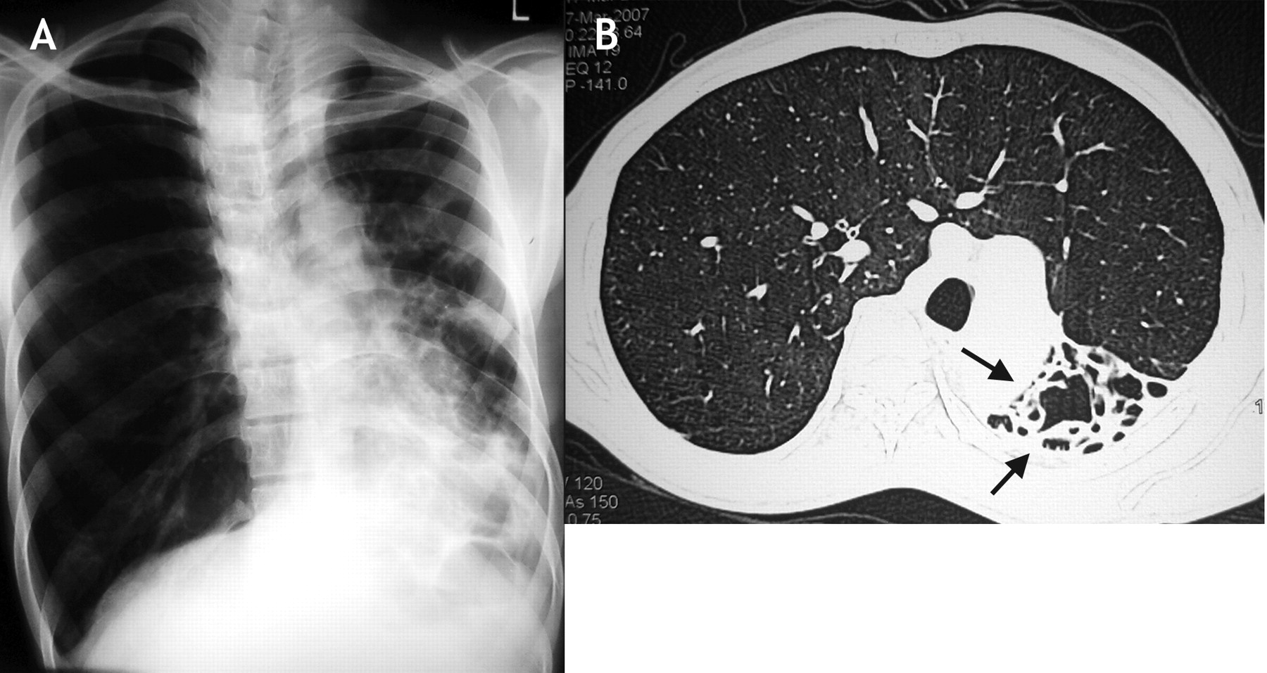
Figure 1: A: Chest radiograph showing volume loss on the left side with bronchiectasis in the left lower lobe and changes on the right consistent with hyperinflation. B: Computed tomography scan of the chest showing the right lung, which has herniated across to the left side in a horseshoe shape, and the destroyed left lung (arrows).

In this issue
Article tools
Respond to this article


Related Articles
Cited By...
- No citing articles found.
More in this TOC Section
Similar Articles
Collections

