


Article Figures & Tables
Figures
Figure 1: Histology image from the placenta showing acute villitis. The villi are distended and contain numerous acute inflammatory cells. There are multiple microabscesses (arrows) with foci of affected villi showing central necrosis (hematoxylin–eosin stain, original magnification × 20)
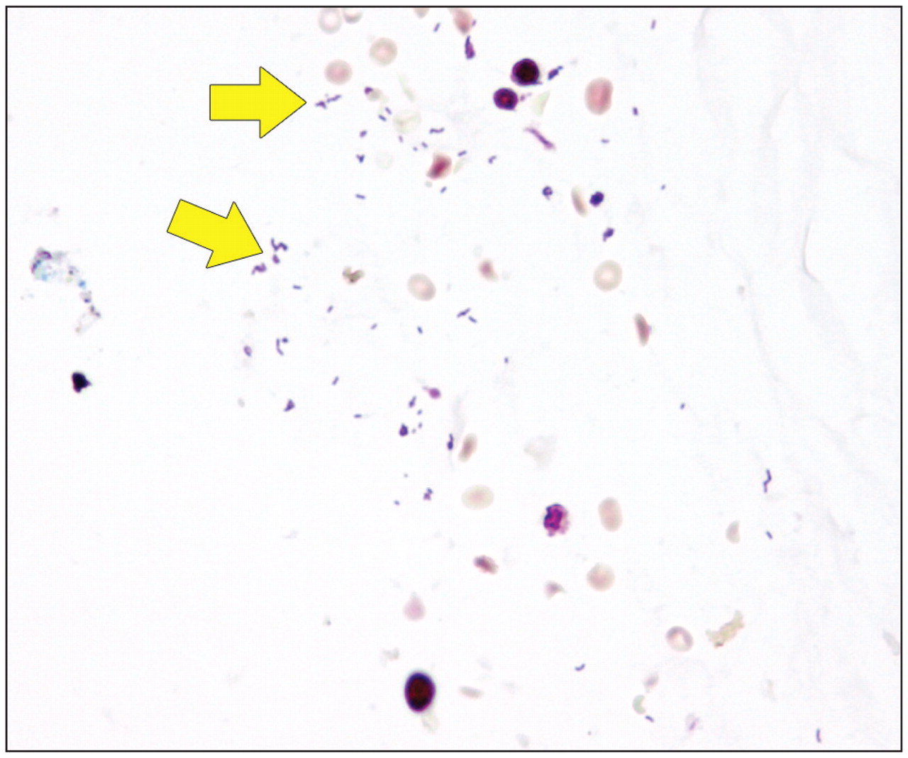
Figure 2: Histology image from the placental membrane showing numerous gram-positive bacilli (arrows) (gram stain, original magnification × 600).


In this issue
Article tools
Respond to this article


Jump to section
Related Articles
Cited By...
- No citing articles found.
More in this TOC Section
Similar Articles

