


Article Figures & Tables
Figures
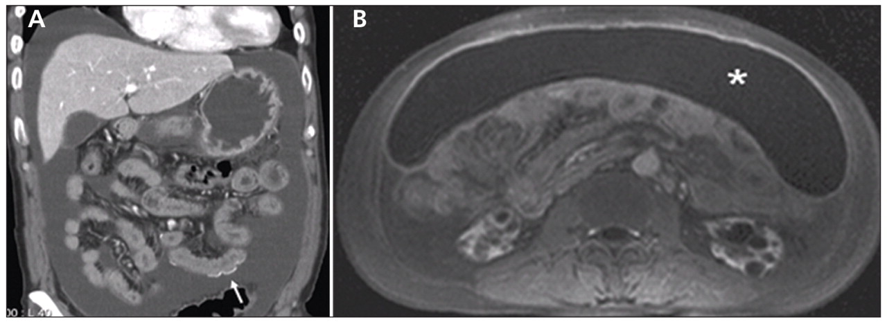
Figure 1: (A) Computed tomography scan of the abdomen showing curvilinear calcifications (arrow) along the bowel walls and serosa. (B) Magnetic resonance imaging scan of the abdomen showing ascites with wall enhancement in the omental space. The small bowel is compressed rather than floating in the ascites.

In this issue
Article tools
Respond to this article


Jump to section
Related Articles
Cited By...
More in this TOC Section
Similar Articles
Collections

