


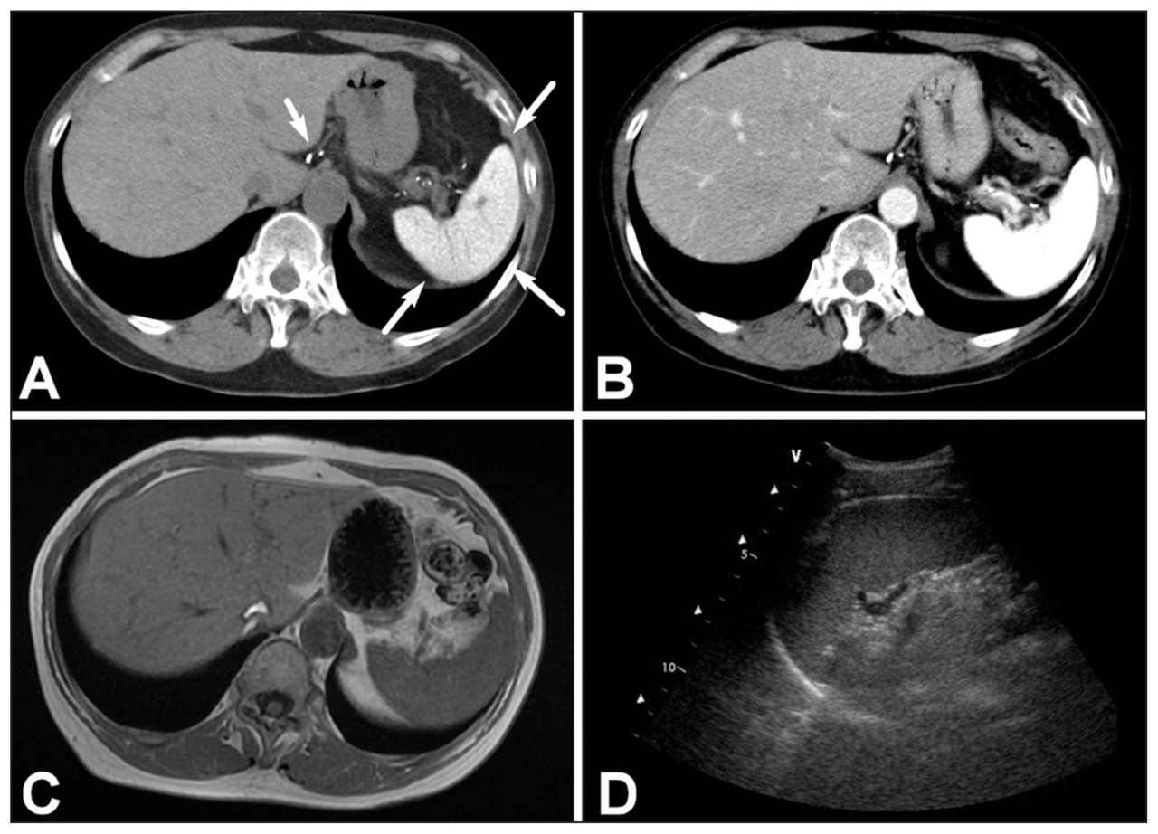
A 60-year-old woman underwent computed tomography imaging of her chest because of a recent episode of hemoptysis. The scan did not show any abnormalities in the thorax. However, unenhanced scans showed a strikingly hyperdense spleen (mean attenuation value 174 [normal 56–65] Hounsfield units) (Figure 1A). There were also hyperdense lymph nodes along the splenoportal axis and faint strands of increased density in the parenchyma of the peripheral liver. Imaging with contrast showed normal size and morphology of the organs with preserved contrast enhancement of the lymph nodes and spleen (Figure 1B).

Unenhanced computed tomography scan of the abdomen (coronal view) of a 60-year-old woman showing strikingly hyperdense spleen and lymph nodes (A); postcontrast images show preserved enhancement of the splenic parenchyma (B); T1-weighted fast spin-echo magnetic resonance image reveals normal signal of liver and spleen (C); ultrasonography of the left hypochondrium shows normal splenic echotexture (D).
Magnetic resonance imaging of the upper abdomen revealed normal signal intensity of spleen and liver both on T1- and T2-weighted images, which ruled out hemochromatosis (Figure 1C). Ultrasonography showed normal splenic echotexture, thereby ruling out calcium deposition (e.g., may occur in autosplenectomy from sickle cell disease) (Figure 1D).
The patient had no history of receiving Thorotrast, a thorium-based angiographic contrast agent, or amiodarone therapy, but she had been given a prolonged course of intramuscular injections of sulfur colloidal gold for rheumatoid arthritis.
The computed tomography findings were consistent with deposition of gold particles in the reticuloendothelial system. Because of gold’s high atomic number, deposition of gold can result in hyperdensity of spleen, liver and lymph nodes on computed tomography imaging.1 Gold, unlike calcium, does not interfere with ultrasound transmission and, unlike iron, is a non-ferromagnetic metal that does not alter the signal intensity of magnetic resonance imaging. Therefore, the ultrasonograph and magnetic resonance images were normal. However, the most important differential diagnosis of a hyperdense spleen is deposition of Thorotrast. This agent was discontinued after the 1950s because of its radioactivity and mutagen risk, which is associated with hepatosplenic malignancies.2 Gold compounds may mimic thorotrastosis at imaging, but their carcinogenic effect is negligible.3
Footnotes
-
This article has been peer reviewed.
-
Competing interests: None declared.
In this issue
Article tools


Jump to section
Related Articles
Cited By...
- No citing articles found.
More in this TOC Section
Similar Articles

