


Article Figures & Tables
Figures
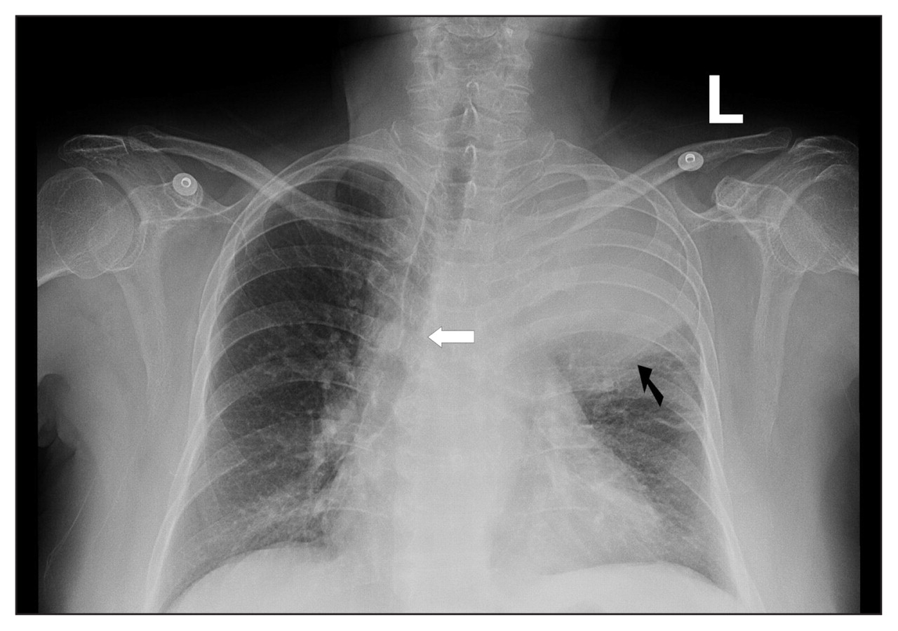
Figure 1: Chest radiograph of a 68-year-old woman showing opacification in the left upper lobe (black arrow) with tracheal displacement to the right (white arrow).
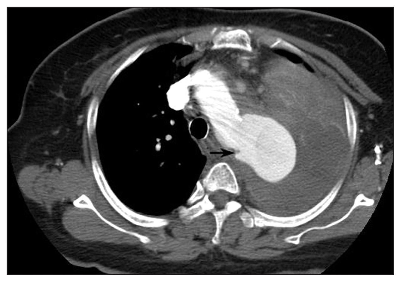
Figure 2: Computed tomography scan of the chest showing a thoracic aortic aneurysm (4 × 6 cm, arrow) with a heterogeneous mass in the left upper lobe consistent with a large hematoma.
In this issue
Article tools
Respond to this article


Jump to section
Related Articles
Cited By...
- No citing articles found.
More in this TOC Section
Similar Articles
Collections

