


Article Figures & Tables
Figures
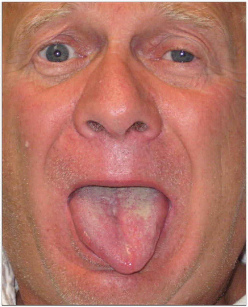
Figure 1: Photograph of a 61-year-old man with intense left-sided headache, showing leftward deviation of the tongue and ptosis and miosis of the left eye.
Box 1: Features of Horner syndrome
Box 2: Signs of medial and lateral medullary syndromes
Figure 2: A: Transverse computed tomography angiography of the neck demonstrates subtle mural soft-tissue thickening of the left internal carotid artery on day two of symptoms (large arrow). B: Transverse T1-weighted magnetic resonance imaging (MRI) with fat saturation on day three of symptoms is consistent with a normal left internal carotid artery (large arrow). C: Transverse T1-weighted MRI with fat saturation on day eight of symptoms demonstrates new mural hyperintensity and thickening of the left internal carotid artery. D: Transverse T1-weighted MRI with fat saturation at six months after presentation shows resolution of the soft-tissue hyperintensity and thickening of the left internal carotid artery. The tortuous but otherwise normal right internal carotid artery is indicated by a small arrow in some of the images.
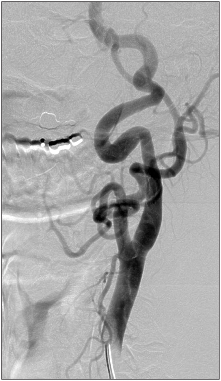
Figure 3: Digital subtraction angiogram after injection of contrast medium into the left common carotid artery on day six of symptoms shows some tortuosity of the vessel but no convincing mural irregularity or luminal narrowing.
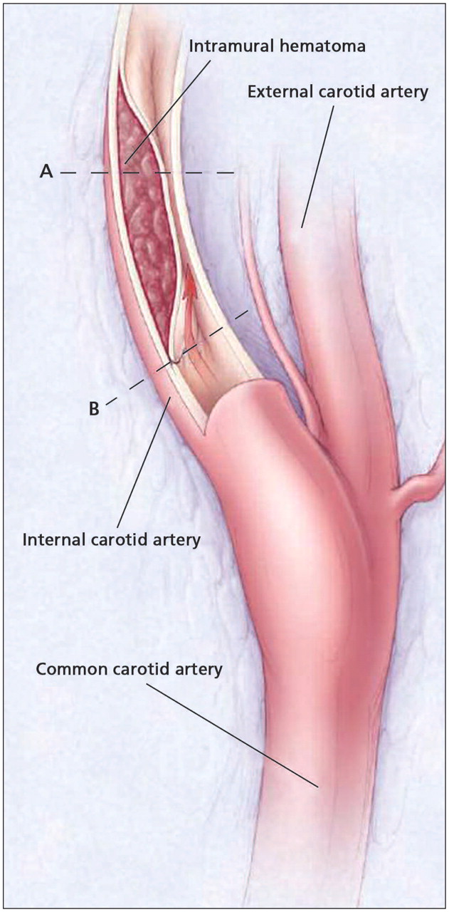
Figure 4: Dissection of the internal carotid artery (site B) causes a mural hematoma to form in the tunica media of the vessel wall (site A). The mural hematoma is causing stenosis of the vessel’s lumen. Reproduced, with permission, from Schievink WI. 1 Spontaneous dissection of the carotid and vertebral arteries. N Engl J Med 2001;344: 898–906. Copyright 2001 Massachusetts Medical Society. All rights reserved.
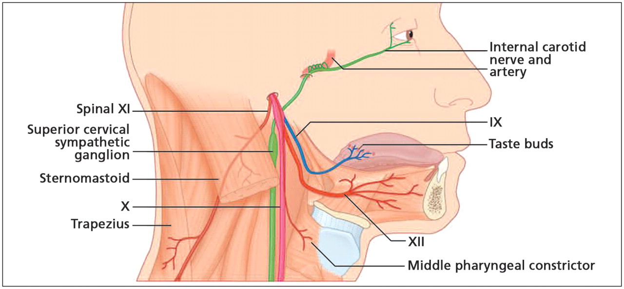
Figure 5: The lowest four cranial nerves are shown emerging from the jugular and hypoglossal foramina, where they join the sympathetic plexus within the carotid sheath. Here, these structures are vulnerable to the compressive effects of a mural hematoma resulting from a carotid dissection. Reproduced, with permission, from FitzGerald MJT, Gruener G, Mtui E. 6Clinical Neuroanatomy and Neuroscience, 5th ed. Figure 18.2. Copyright 2007 Elsevier.


In this issue
Article tools


Related Articles
Cited By...
More in this TOC Section
Similar Articles
Collections

