


Article Figures & Tables
Figures
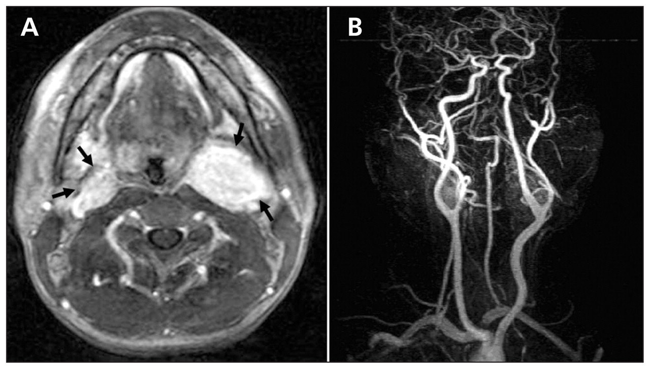
- Figure 1:
(A) Axial T1-weighted magnetic resonance (MR) image, with gadolinium enhancement, of the neck of a 42-year-old man showing bilateral, well-circumscribed masses (arrows) with a “salt and pepper” appearance; masses measured 2.0 × 1.6 cm (right) and 4.0 × 3.3 cm (left). (B) MR angiogram showing splaying of external and internal carotid arteries (lyre sign), bilaterally.
In this issue
Article tools
Respond to this article


Related Articles
Cited By...
- No citing articles found.
More in this TOC Section
Similar Articles
Collections

