


An otherwise healthy 71-year-old woman presented to our clinic with a history of pruritus and burning of the vulva. On examination, we observed an atrophic, white, shiny plaque involving the vulva, perineum and perianal areas. The clitoral hood and labia minora were completely scarred and the introitus was narrow (Figure 1). We diagnosed vulvar lichen sclerosus, a chronic inflammatory disease affecting the anogenital area that can present in any age group, though it is most common before puberty and after menopause.1,2 We noted no areas of concern for malignant disease, so we did not take a biopsy. We treated the patient with high-potency steroids (clobetasol ointment twice daily for 6 wk, then daily). She responded well to treatment, and once her disease settled, we switched her to mid-potency steroids (betamethasone valerate 0.1% ointment daily).
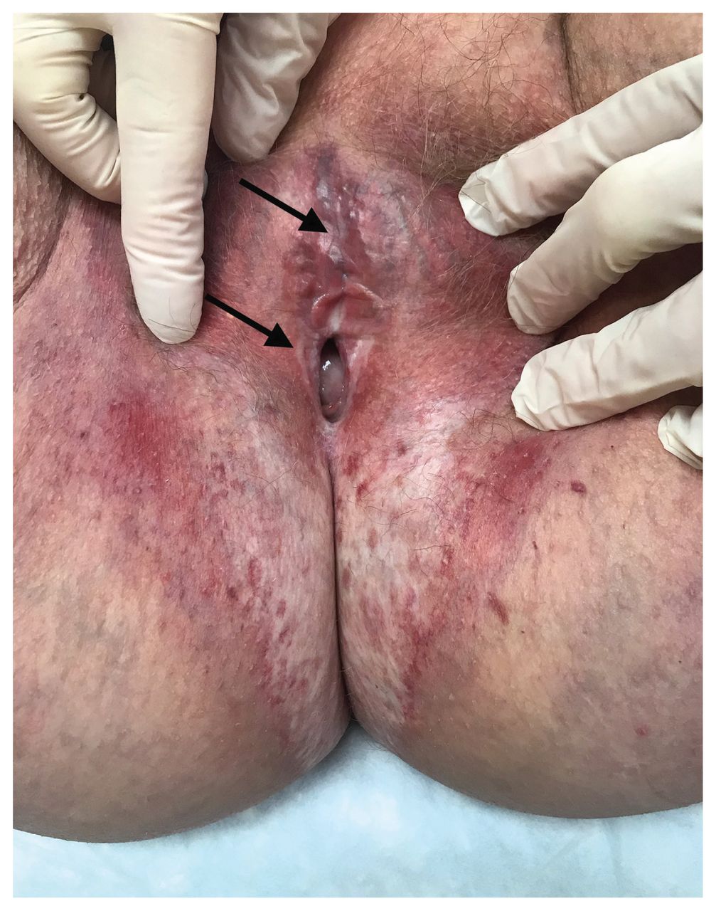
Photograph of a 71-year-old woman with vulvar lichen sclerosus, showing a large, atrophic, shiny, white plaque with petechia, involving the vulva, perineum and perianal areas in the classic “figure of eight” distribution. Severe scarring and disappearance of the clitoral hood and labia minora bilaterally (arrows), as well as substantial narrowing of the introitus, are evident.
The classic clinical features of vulvar lichen sclerosus are shiny white atrophic plaques in a “figure of 8” pattern that encircle the vulva, perineum and perianal areas, although more focal areas can be affected.1,2 Advanced disease can result in severe scarring, leading to deformed anatomy and stenosis. Other findings may include petechia, ecchymosis, erosions, fissures, hypertrophic plaques and hyperpigmentation. Fifteen percent of patients will also have extragenital involvement.1–3 Vulvar lichen sclerosus is rarely asymptomatic. More than 90% of patients will present with severe pruritus, but they may also have dysuria, dyspareunia and clitoral hyperesthesia. Differential diagnoses include lichen planus, dermatitis or lichen simplex chronicus. In children, sexual abuse must always be excluded.1,3
Therapy is administered in 2 phases: an ointment of ultrapotent topical corticosteroids (e.g., clobetasol propionate 0.05%) to induce remission, followed by long-term maintenance therapy with daily topical corticosteroids of moderate potency (e.g., betamethasone valerate 0.1%) or twice weekly ultrapotent ointments. Many studies have shown the excellent safety profile of long-term topical corticosteroids in the mucosal vulvar area.1–3 Treating vulvar lichen sclerosus is critical to control symptoms, prevent scarring and decrease the risk of associated squamous cell carcinoma.2 If inadequately treated, patients have a 50% risk of scarring and a 3%–5% risk of malignant transformation. Biopsy is not required to make a diagnosis of vulvar lichen sclerosus; however, failure to improve on treatment should prompt biopsy to rule out squamous cell carcinoma. Regular follow-up is encouraged to optimize treatment compliance, adjust potency of topical corticosteroids and monitor for complications.1,3
Footnotes
Competing interests: None declared.
This article has been peer reviewed.
The authors have obtained patient consent.
This is an Open Access article distributed in accordance with the terms of the Creative Commons Attribution (CC BY-NC-ND 4.0) licence, which permits use, distribution and reproduction in any medium, provided that the original publication is properly cited, the use is noncommercial (i.e., research or educational use), and no modifications or adaptations are made. See: https://creativecommons.org/licenses/by-nc-nd/4.0/
In this issue

Article tools


Jump to section
Related Articles
Cited By...
More in this TOC Section
Similar Articles
Collections

