


- © 2007 Canadian Medical Association
What's your call?

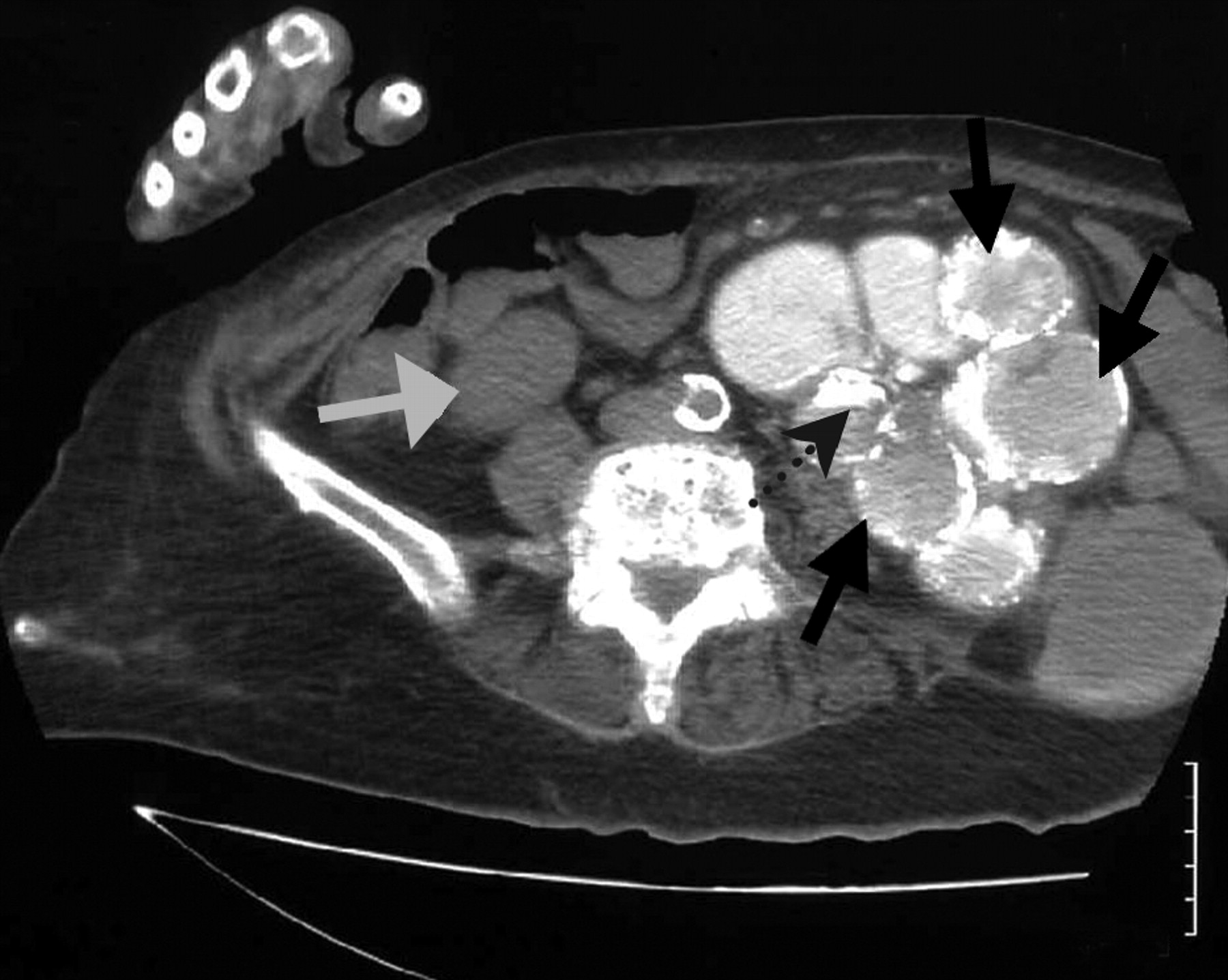
Computed tomography scan of the abdomen of an 87-year-old woman with 4-day history of fever, dysuria and left-flank pain.
This patient had a history of renal colic and recurrent urinary tract infections. On physical examination she had tenderness at her left costophrenic angle. She had pyuria on a routine urinalysis, leukocytosis (leukocyte count 15.6 × 10⁹ /L, 0.88 neutrophils) and acute renal failure (serum creatinine level 574.6 μmol/L). Computed tomography of the abdomen showed a nonfunctioning left kidney and radiographic findings compatible with xanthogranulomatous pyelonephritis (Figure 1). (Additional computed tomography scans showing the extent of the patient's kidney disease can be seen in Appendix 1, available at www.cmaj.ca/cgi/content/full/177/9/1027/DC1.)

Figure 1: Computed tomography scan of the abdomen showing renal stone (dashed arrow) and cystic lesions (black arrows). Low pole of the normal right kidney is indicated with the grey arrow.
Escherichia coli grew in a urine culture. The patient was prescribed broad-spectrum antibiotics (piperacillin and tazobactam), with significant clinical improvement. She was discharged 10 days later.
Xanthogranulomatous pyelonephritis is a severe, chronic bacterial infection of the kidneys characterized by the destruction of the renal parenchyma and the presence of granulomas, abscesses and foam cells.
Footnotes
-
Competing interests: None declared.
In this issue

Article tools


Jump to section
Related Articles
Cited By...
- No citing articles found.
More in this TOC Section
Similar Articles
Collections

