


Article Figures & Tables
Figures
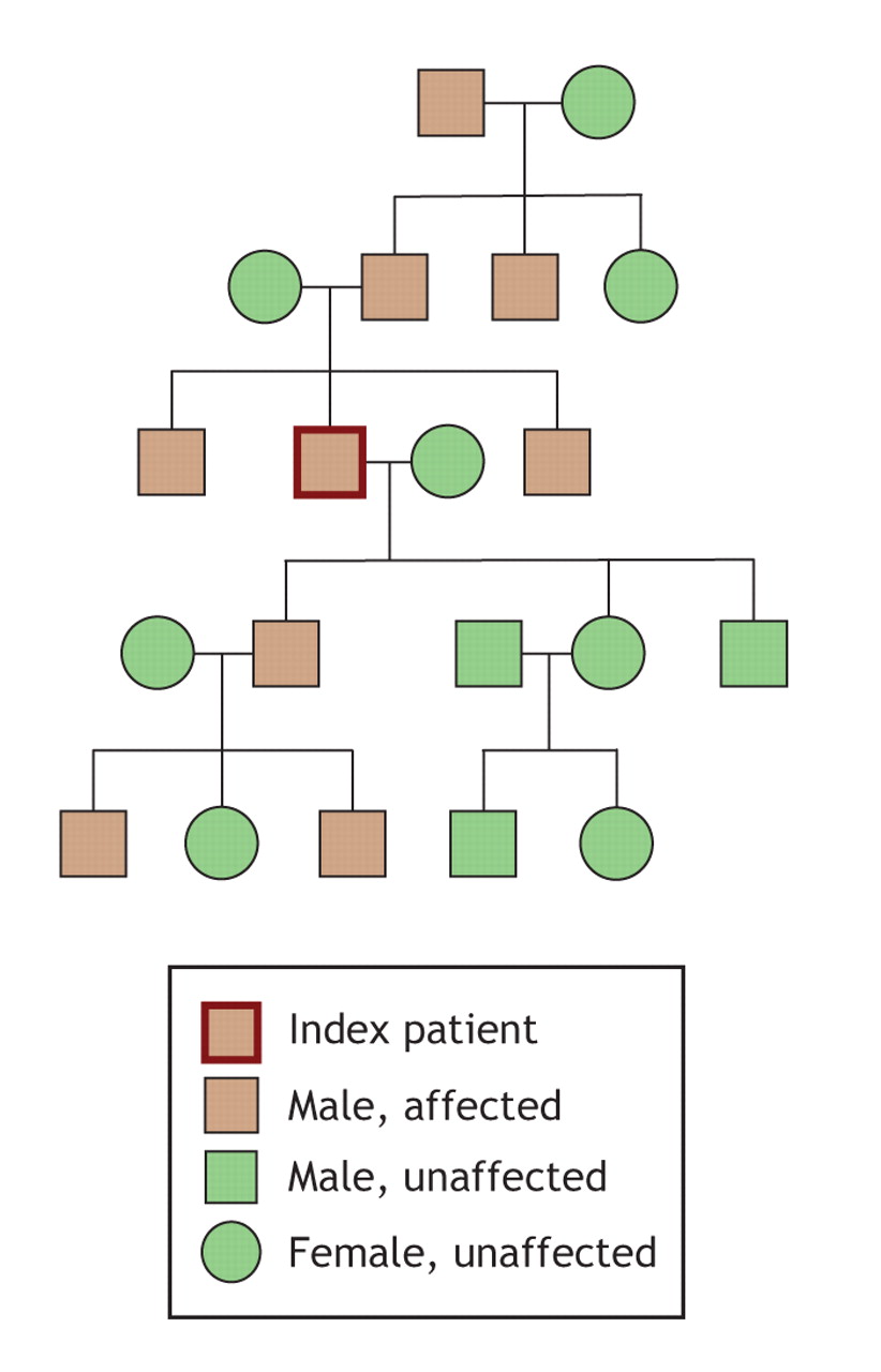
Fig. 1: Pedigree of the patient's affected family members (brown boxes; those not affected are shown in green).
Fig. 2: Photograph of the patient's knees, showing bilateral bony outgrowths around both joints and a genu valgum on the left leg only.
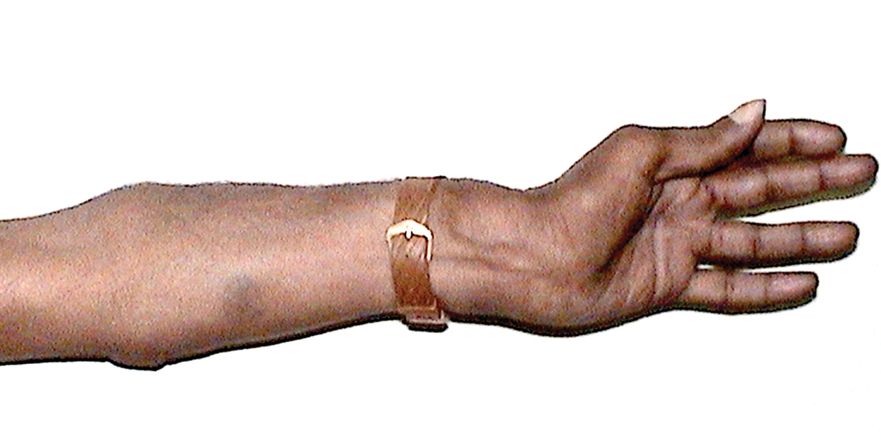
Fig. 3: Ulnar deviation of the patient's left forearm.
Fig. 4: Chest radiograph (left) and CT scan revealing a persistent lesion (exostosis) on the patient's left fourth rib.
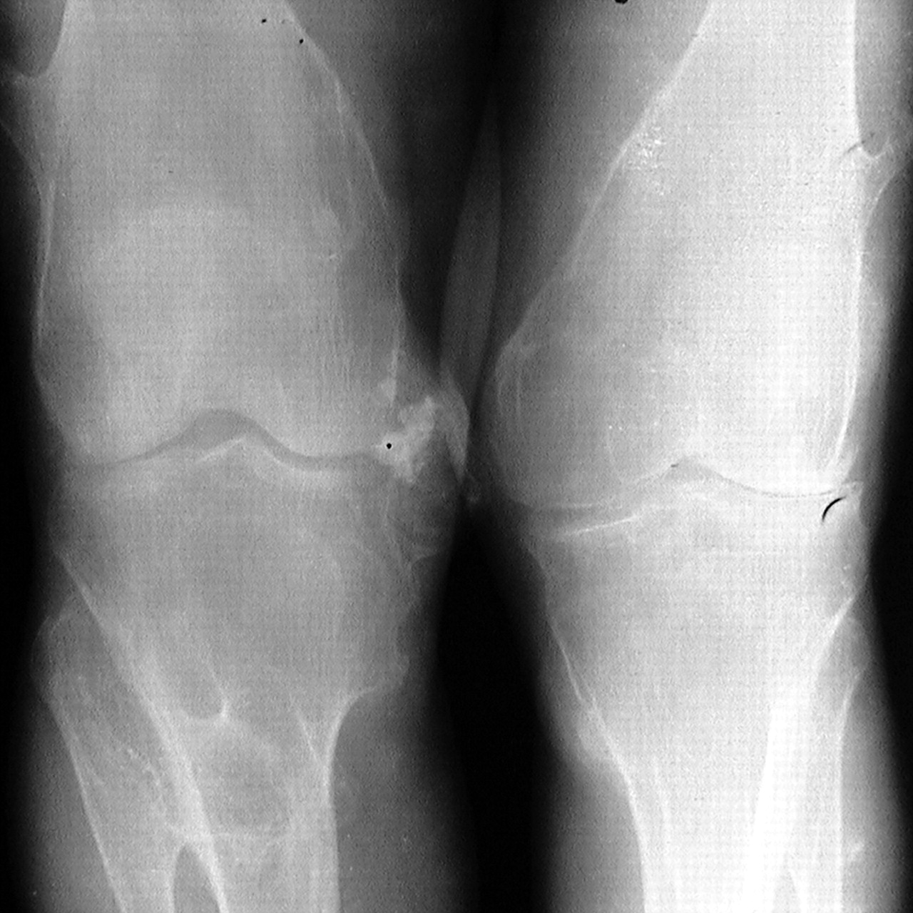
Fig. 5: Radiograph of the knee region; bilateral bony outgrowths are evident.
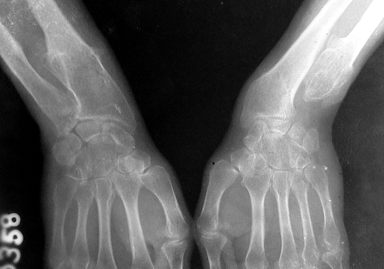
Fig. 6: Radiograph of the patient's wrists. Note the lesion at the lower end of the left ulna.


In this issue
Article tools
Respond to this article


Jump to section
Related Articles
Cited By...
- No citing articles found.
More in this TOC Section
Similar Articles

