


Article Figures & Tables
Figures
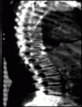
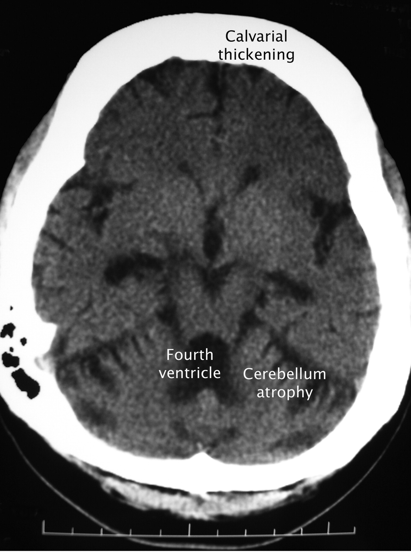
Figure. Axial plain CT scan of a 31-year-old woman with long-standing epilepsy who injured her head during a seizure.
Fig. 1: Axial plain CT scan after prolonged anticonvulsant therapy showing cerebellar atrophy with secondary dilation of the fourth ventricle and marked (symmetrical) hyperostosis of the skull.


In this issue
Article tools
Respond to this article


Jump to section
Related Articles
Cited By...
- No citing articles found.
More in this TOC Section
Similar Articles

